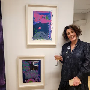
I eventually got to see it!!!
And to tell you I was shocked when I visited Lough Neagh this time is an understatement!!! I visited a different part of the Lough this time by the Boathouse on the the Lough Shore Road, Antrim. Even though I had seen many images of it on social media, I still wasn’t prepared for what I saw- the colour and the smell…I could actually smell it as I drove closer-it smells like bad rotting garbage or sewage and seemingly the smell gets worse as it gets older and decays.
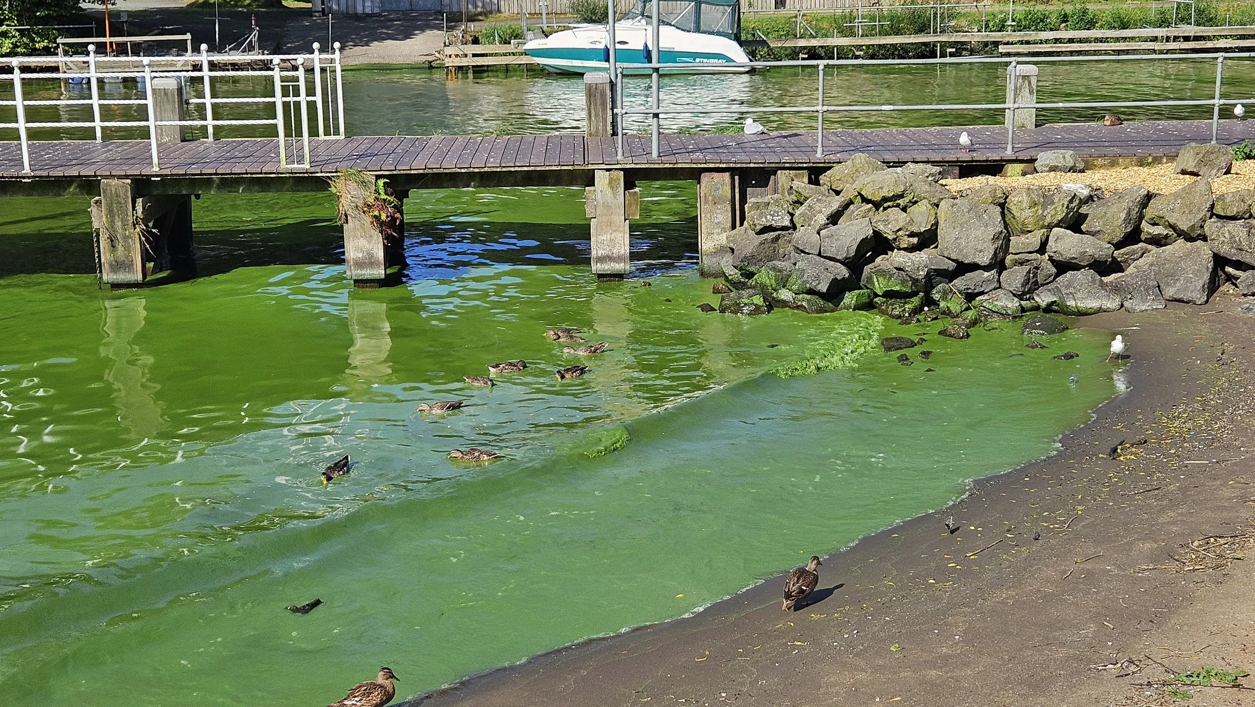
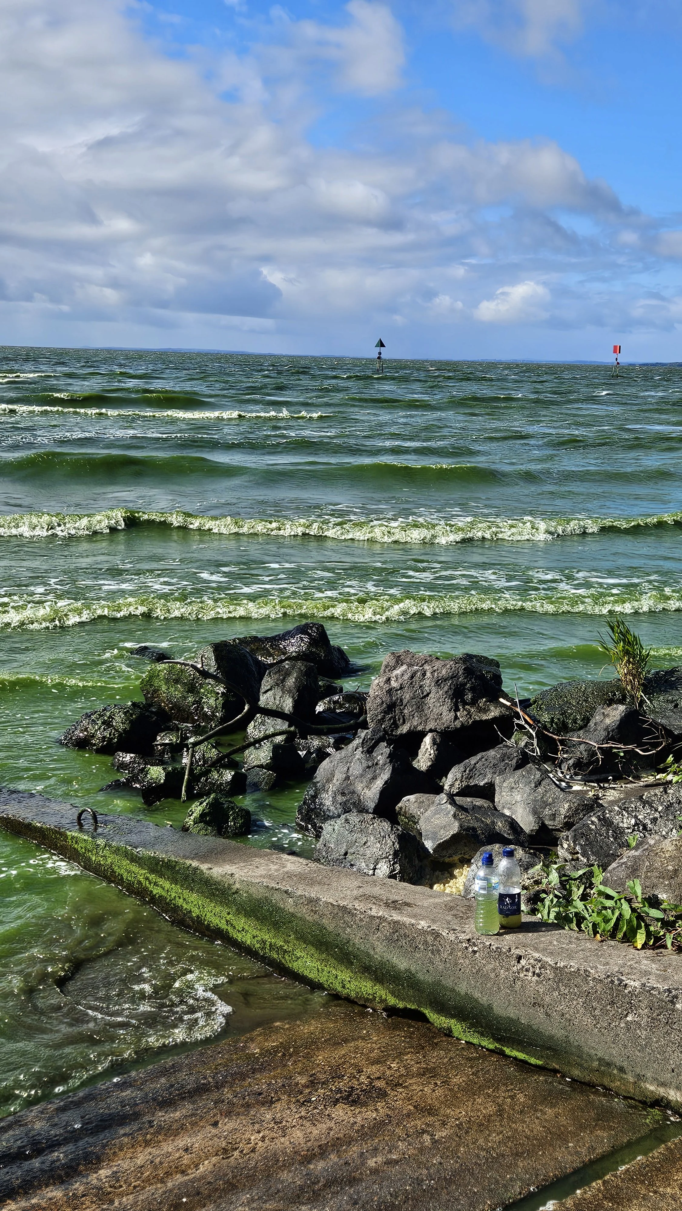
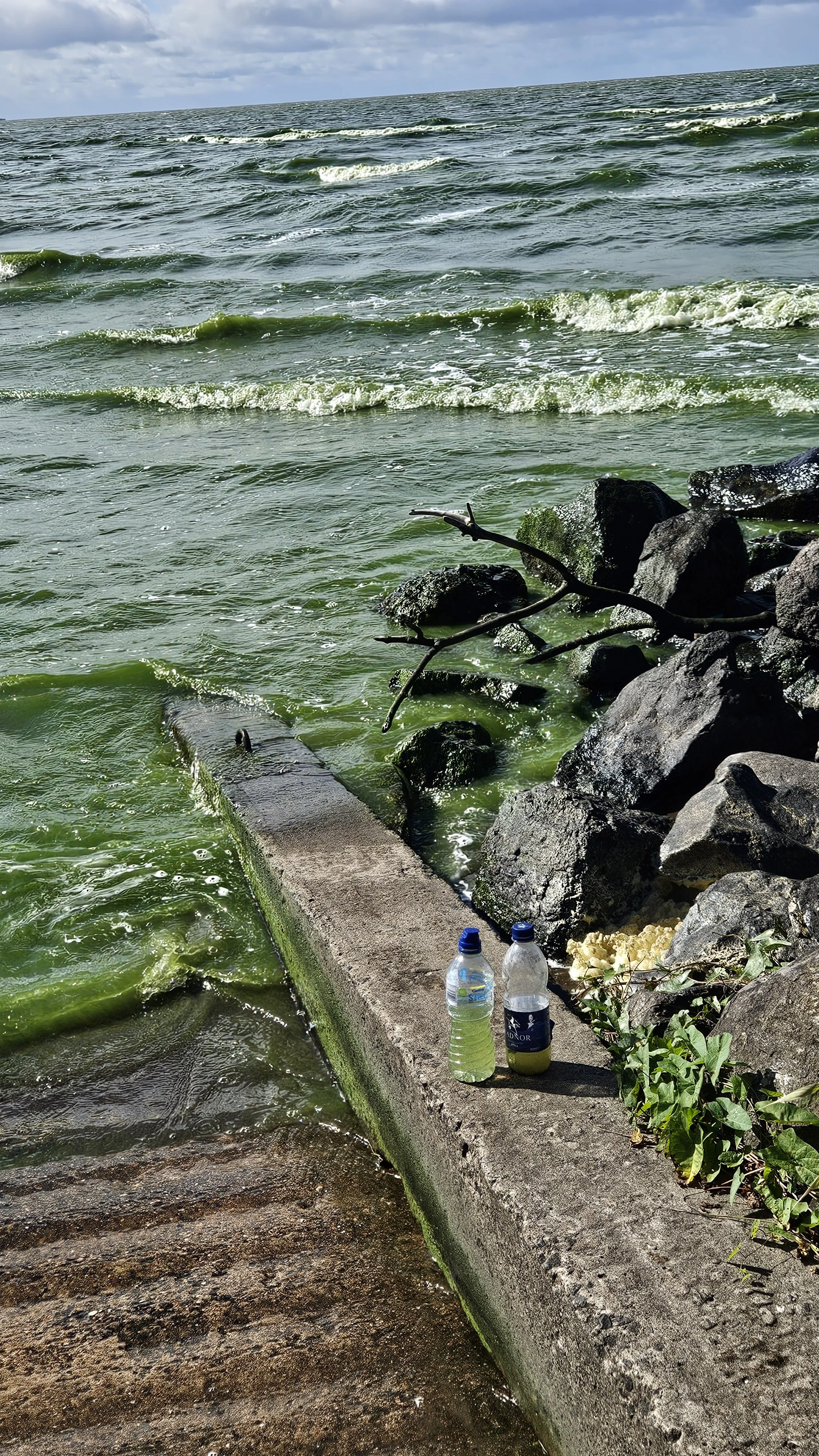
As I walked around the shore, there was something post-apocalyptic about it and a strange green residue had settled over the shoreline and everything was eerily quiet. It reminded me of that scene in the Planet of the Apes (the original- coz I am old lol…)when the Statue of Liberty was buried in the sand.
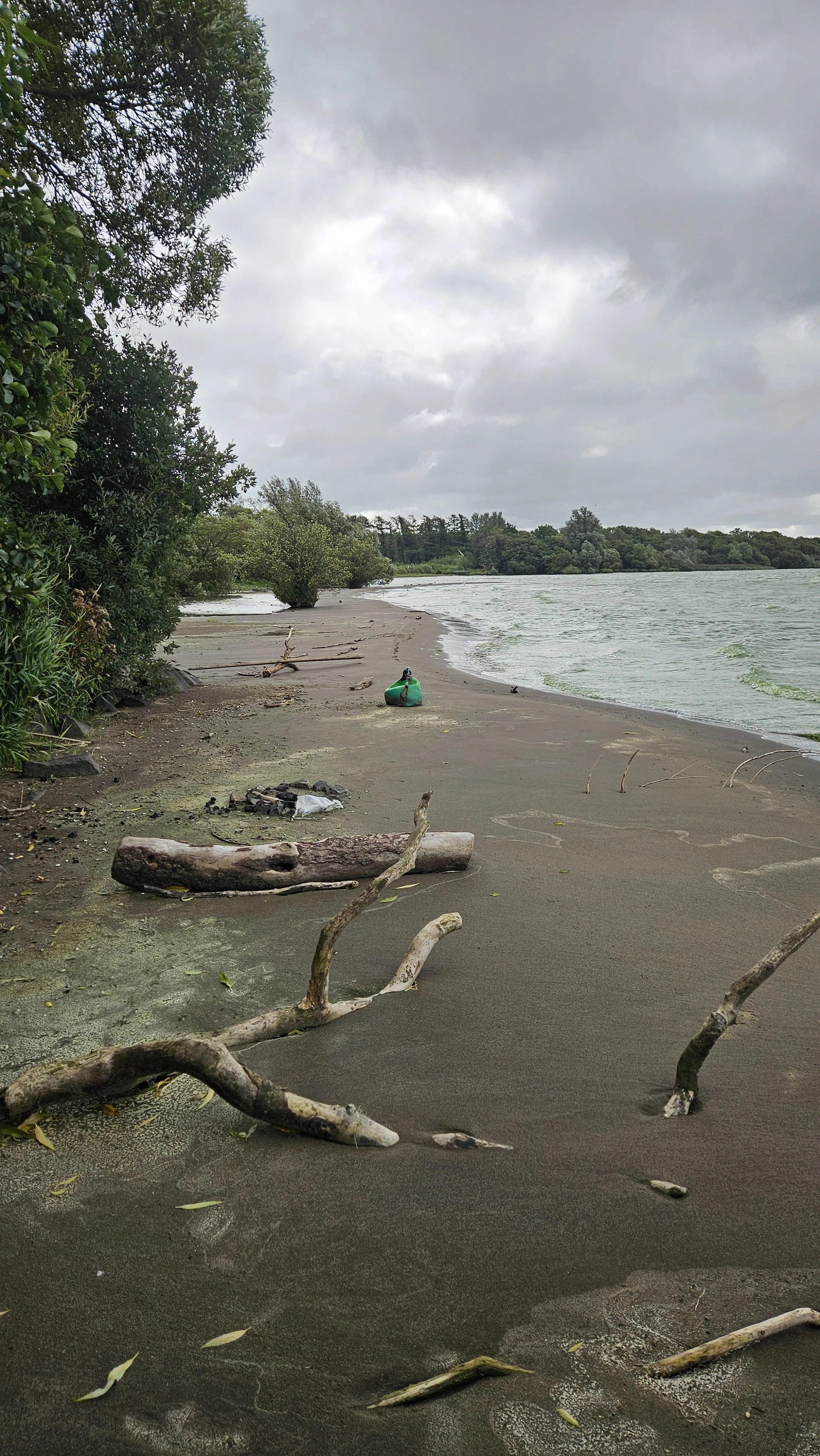
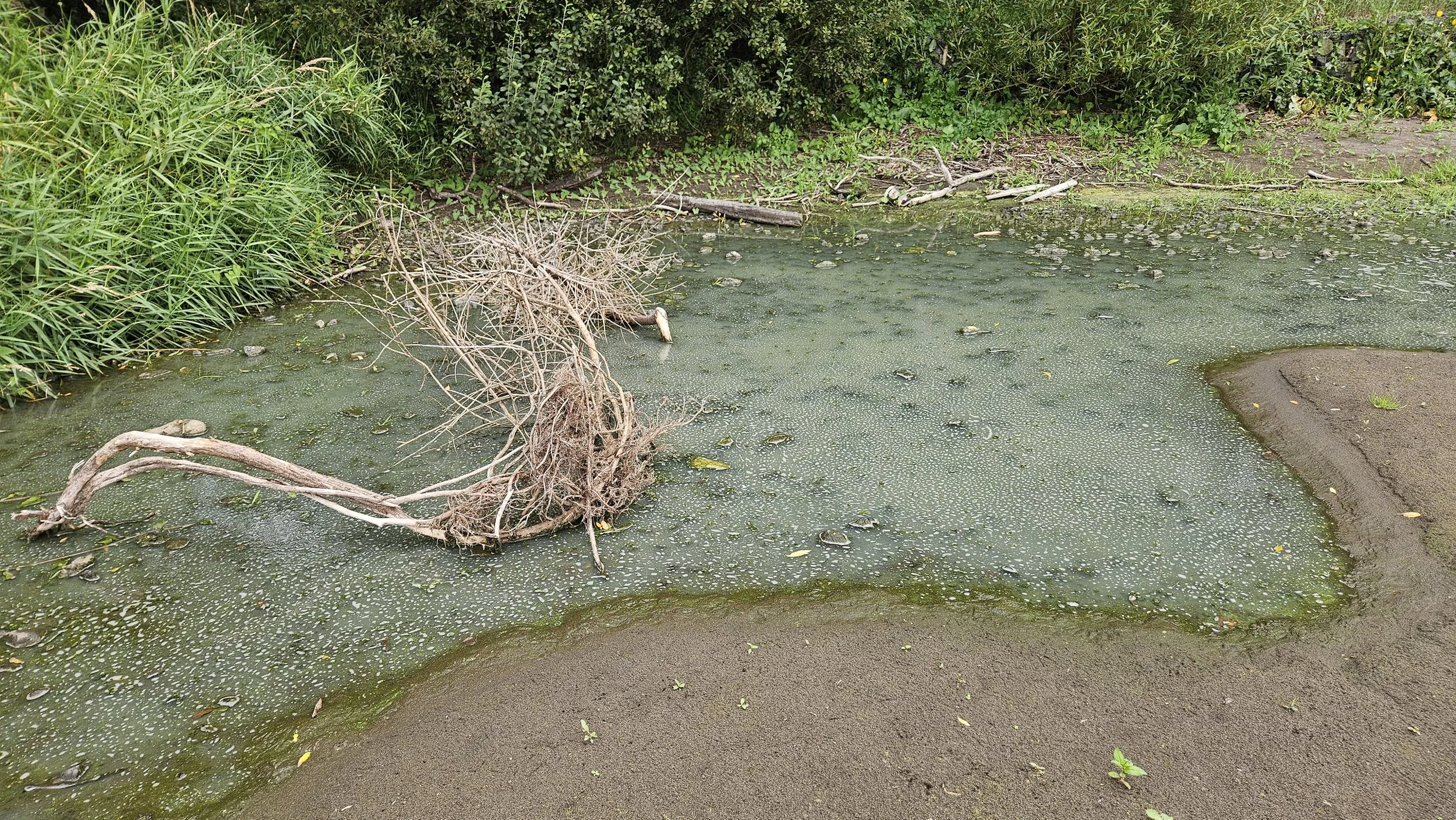
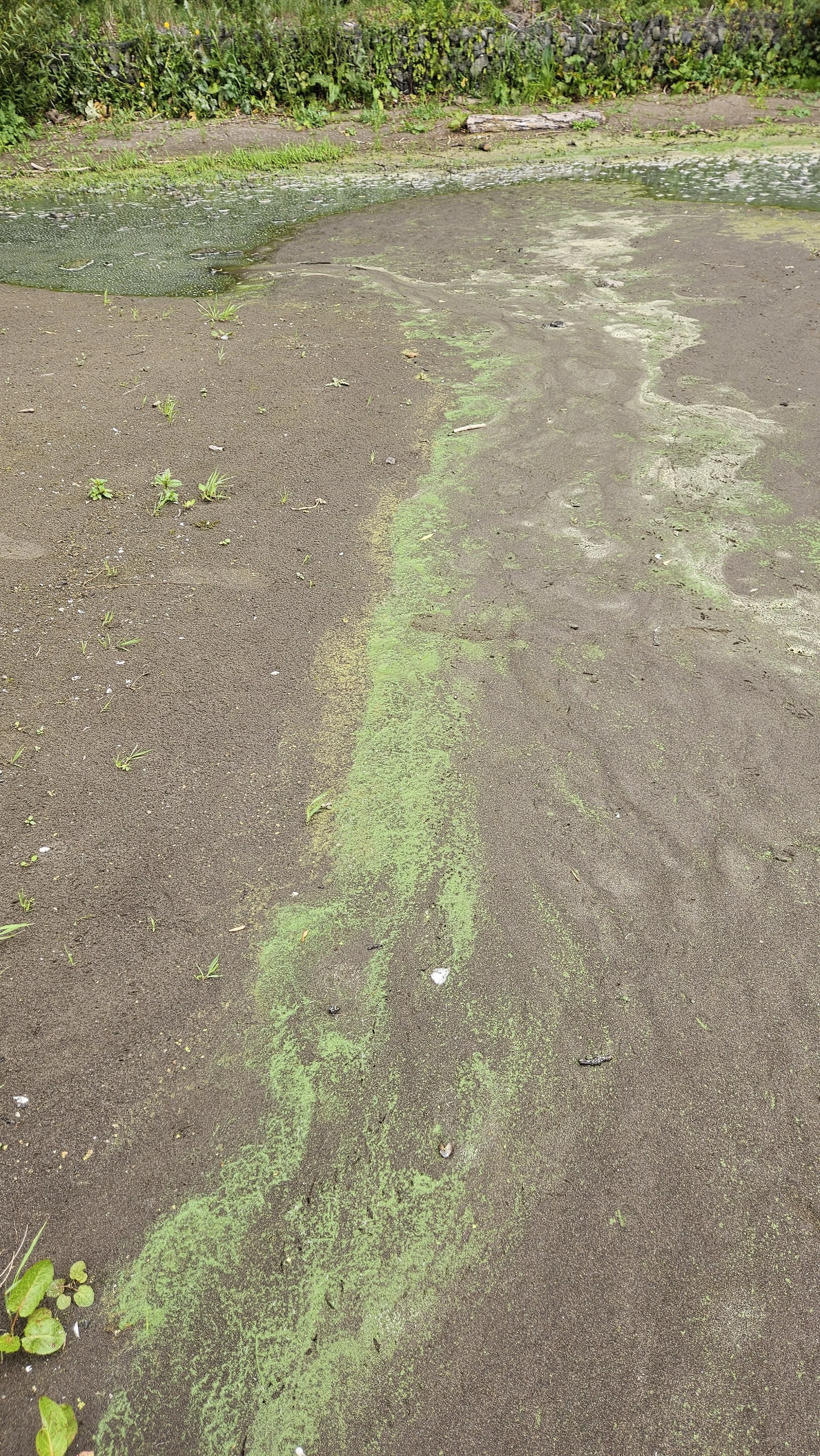
I took samples of the algae filled water and sediment from above and the smell off the bottles was unreal driving home…
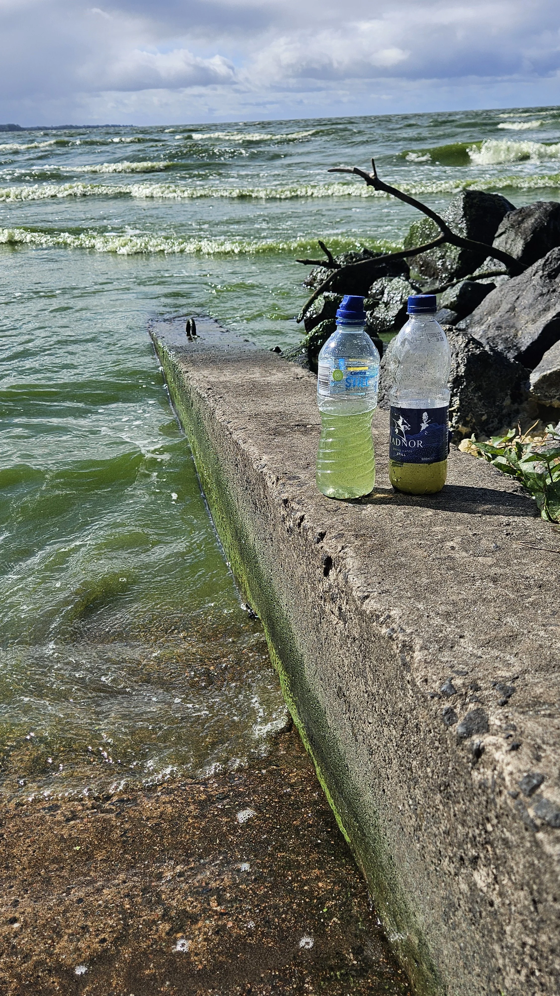
Samples of water from Lough Neagh and sediment from the shoreline
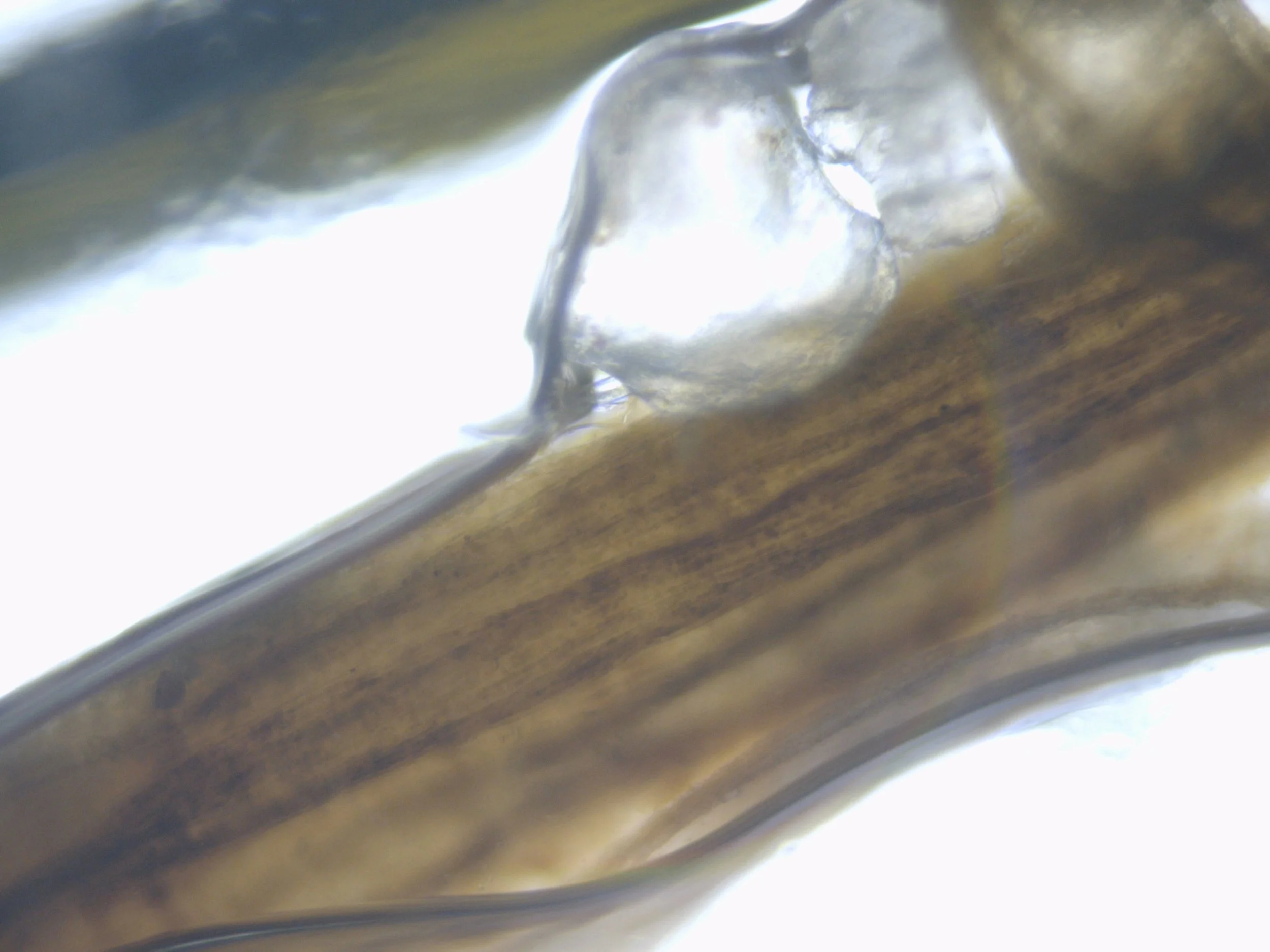
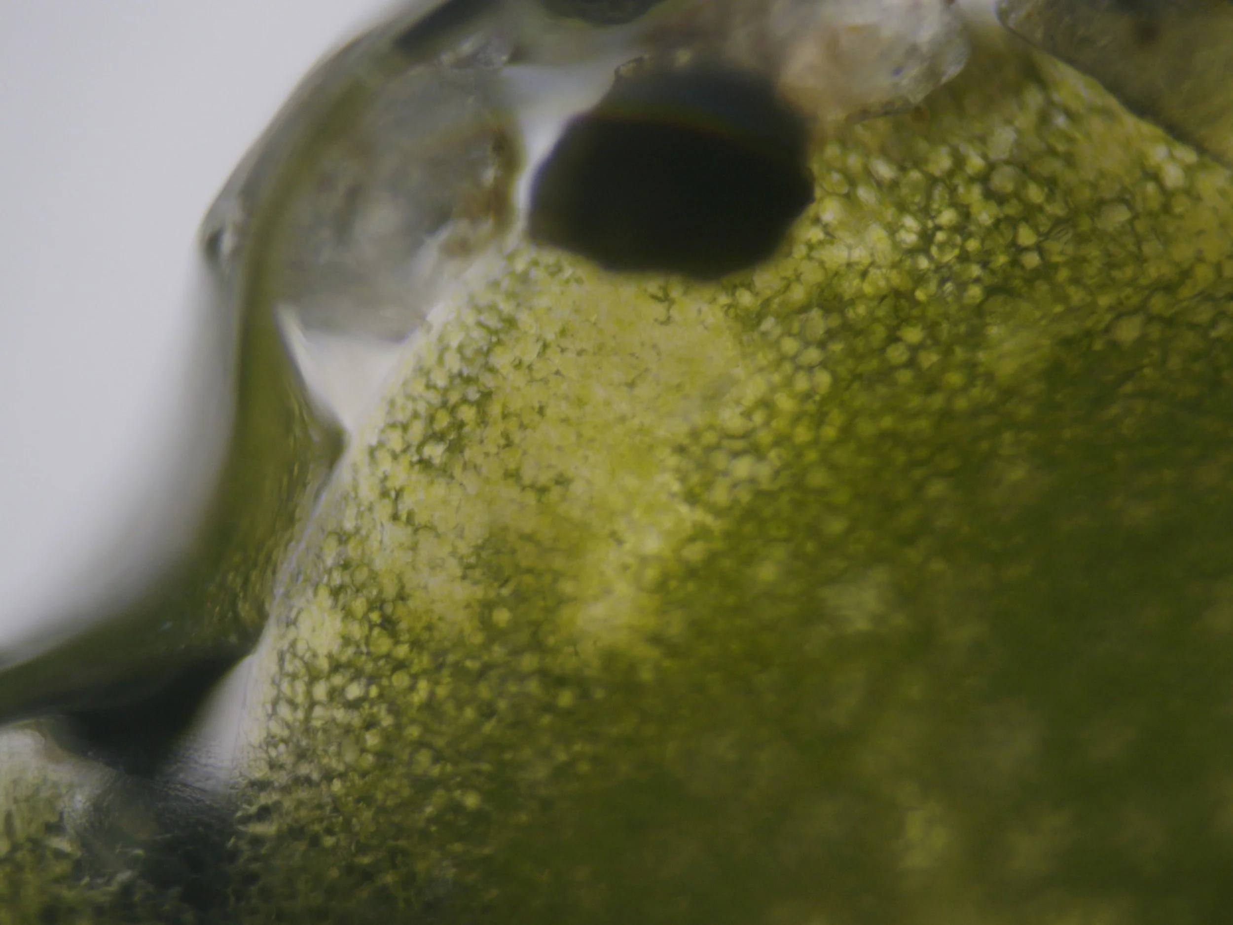
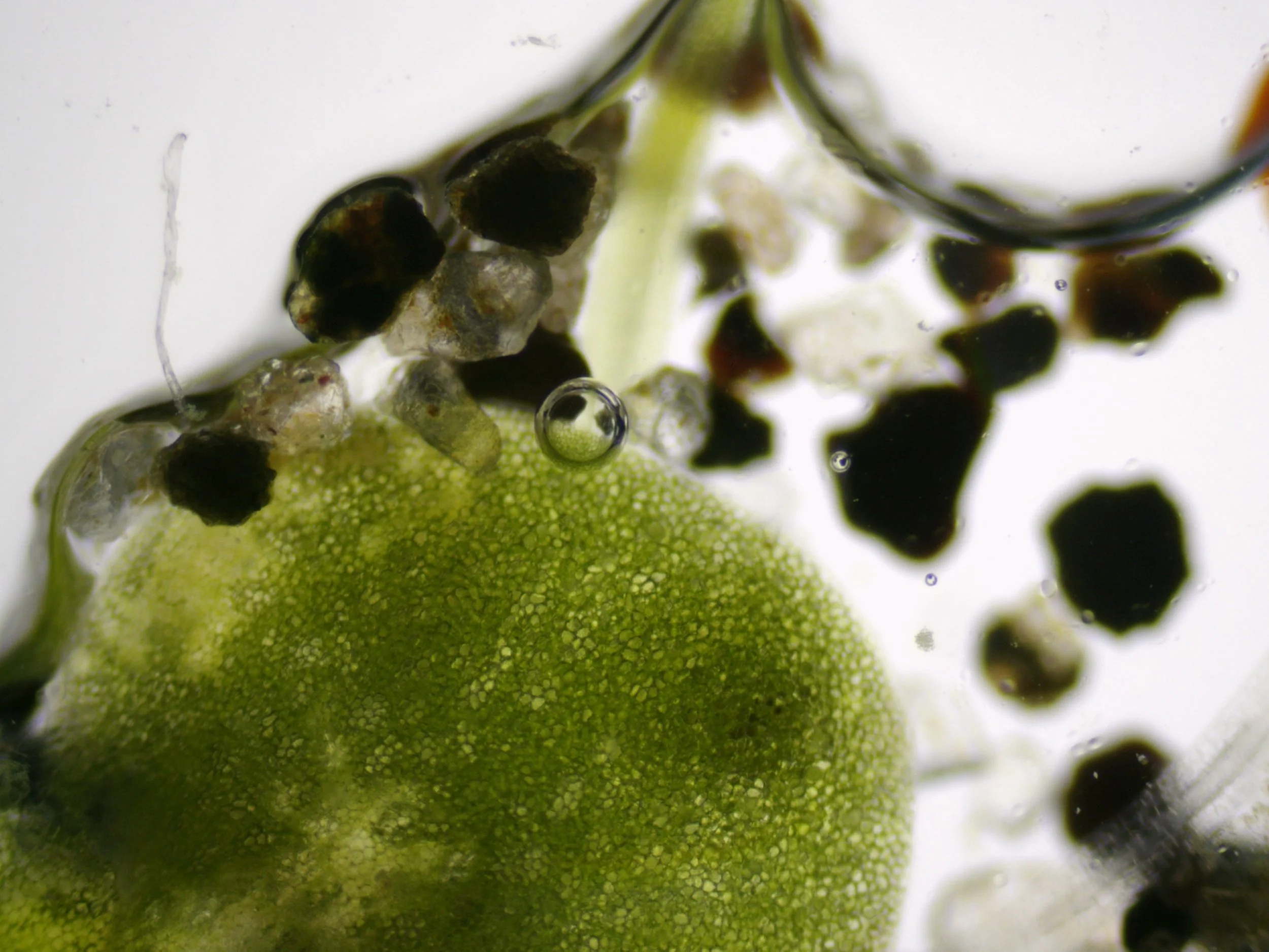
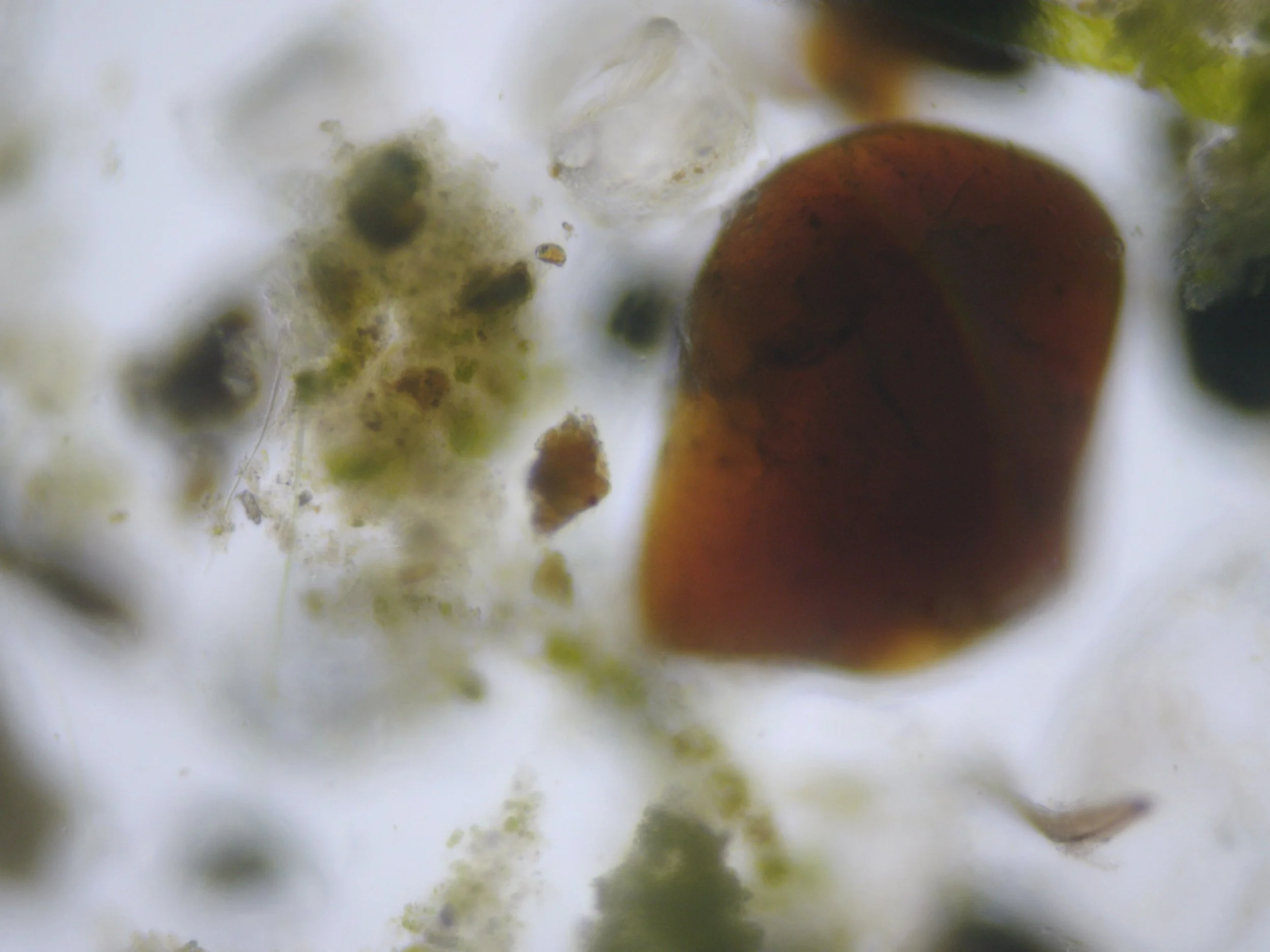
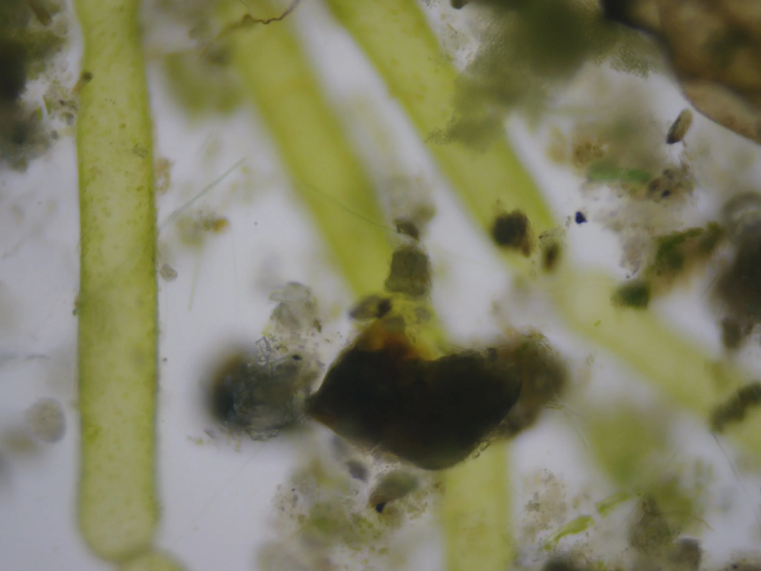
Before I used the microscopic stains,the Lough Neagh samples appeared surprisingly muted under the microscope. Much of the material was translucent and low in contrast, making individual structures difficult to distinguish from the surrounding water and sediment. These unstained images became an important baseline, however, allowing me to compare what was already present in the sample with what became visible after the introduction of Methylene Blue and Lugol's Iodine.
The main reason is for that is actually very simple: most biological material is surprisingly transparent under ordinary bright-field microscopy. Without a stain, there often isn't enough contrast between the structures in the specimen and the surrounding water. Cell walls, membranes and internal structures can all have quite similar optical properties, so they tend to merge visually rather than leap out at you.
Unstained samples below:
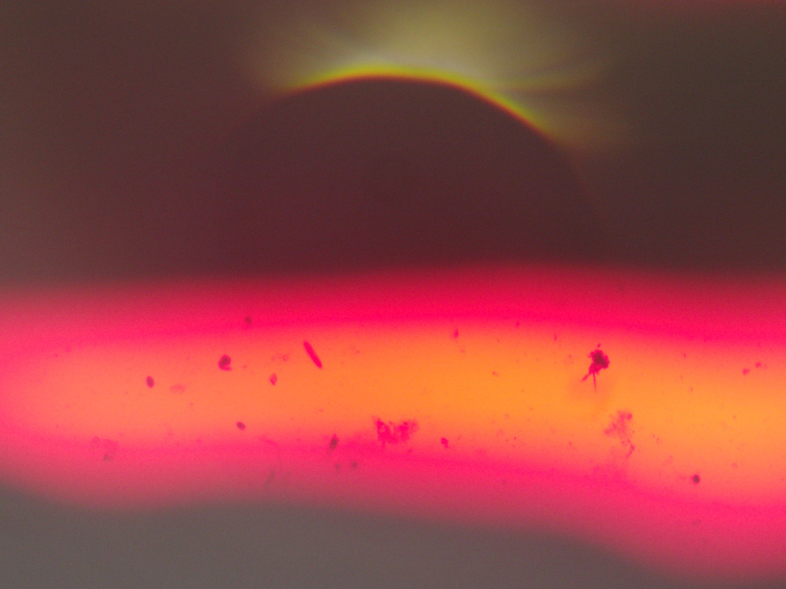
Using Lugols Iodine stain below…
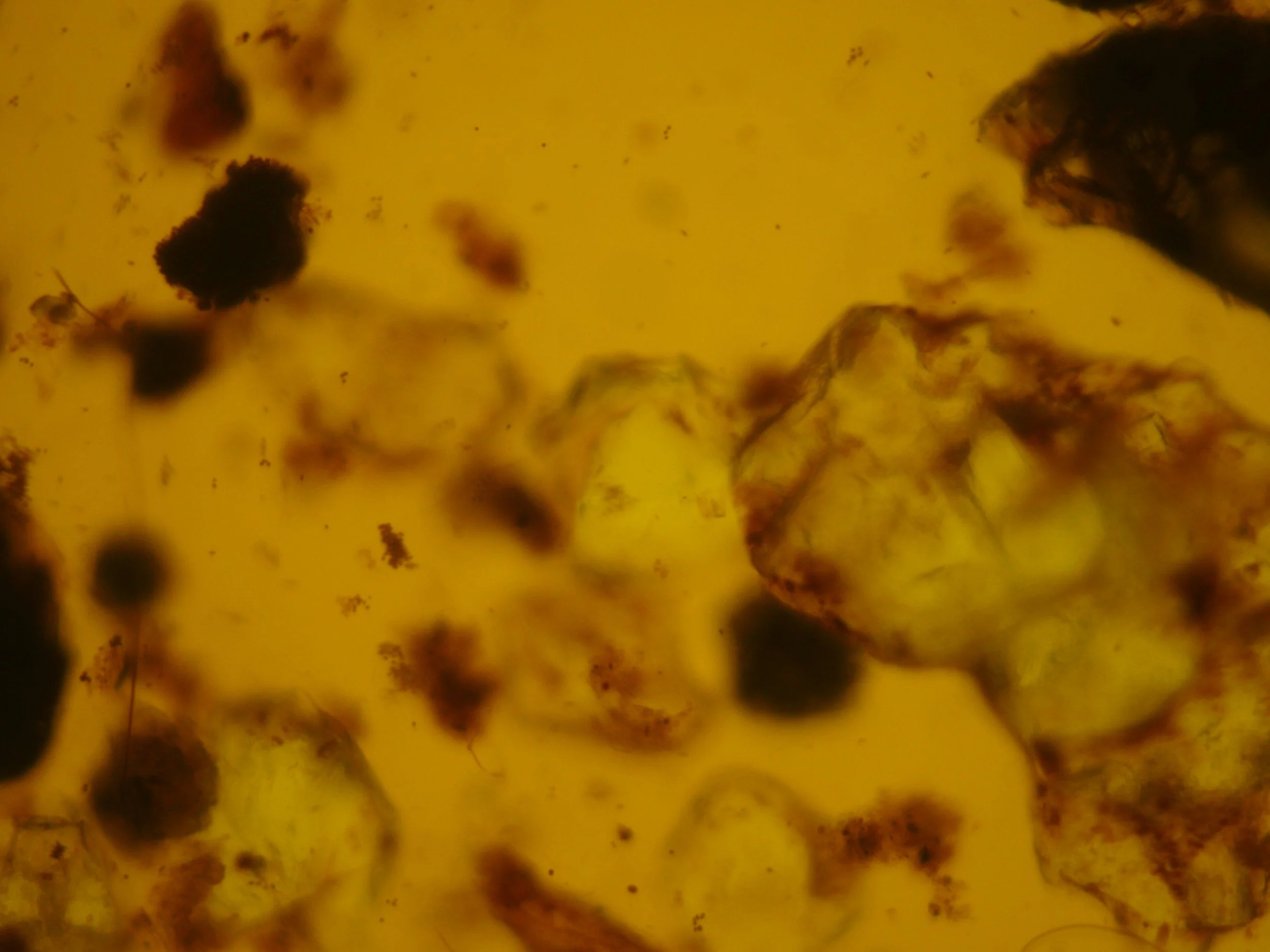
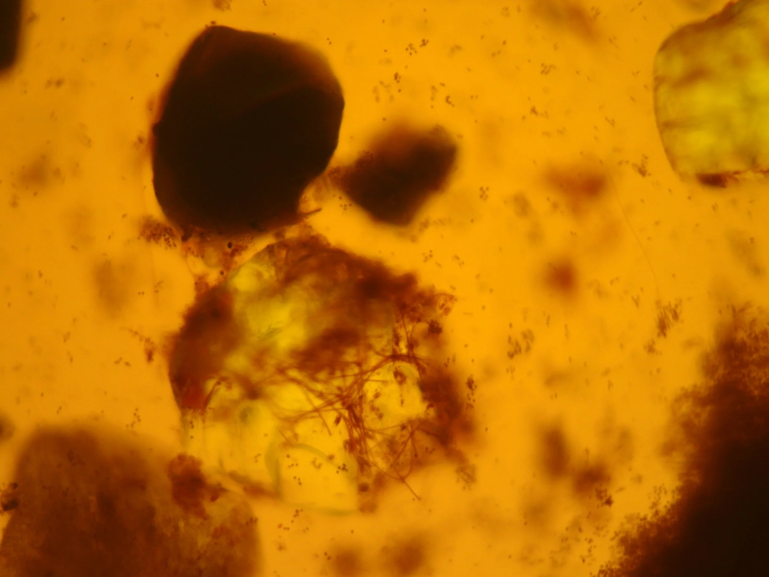
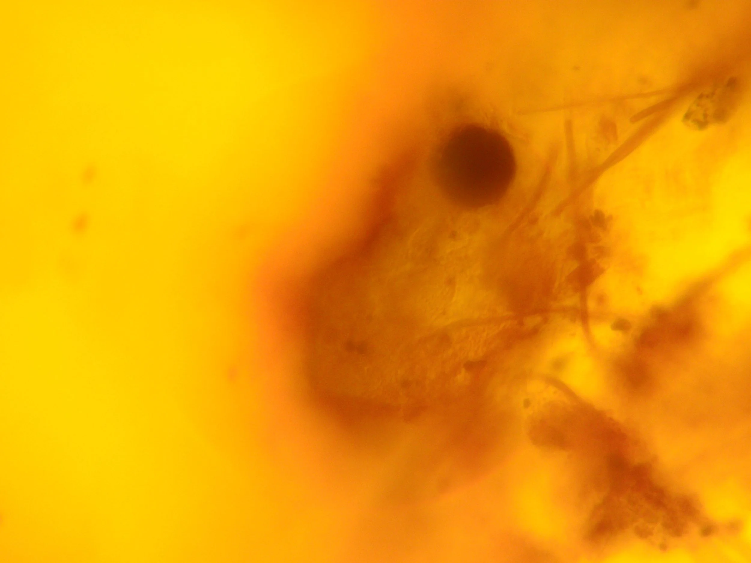
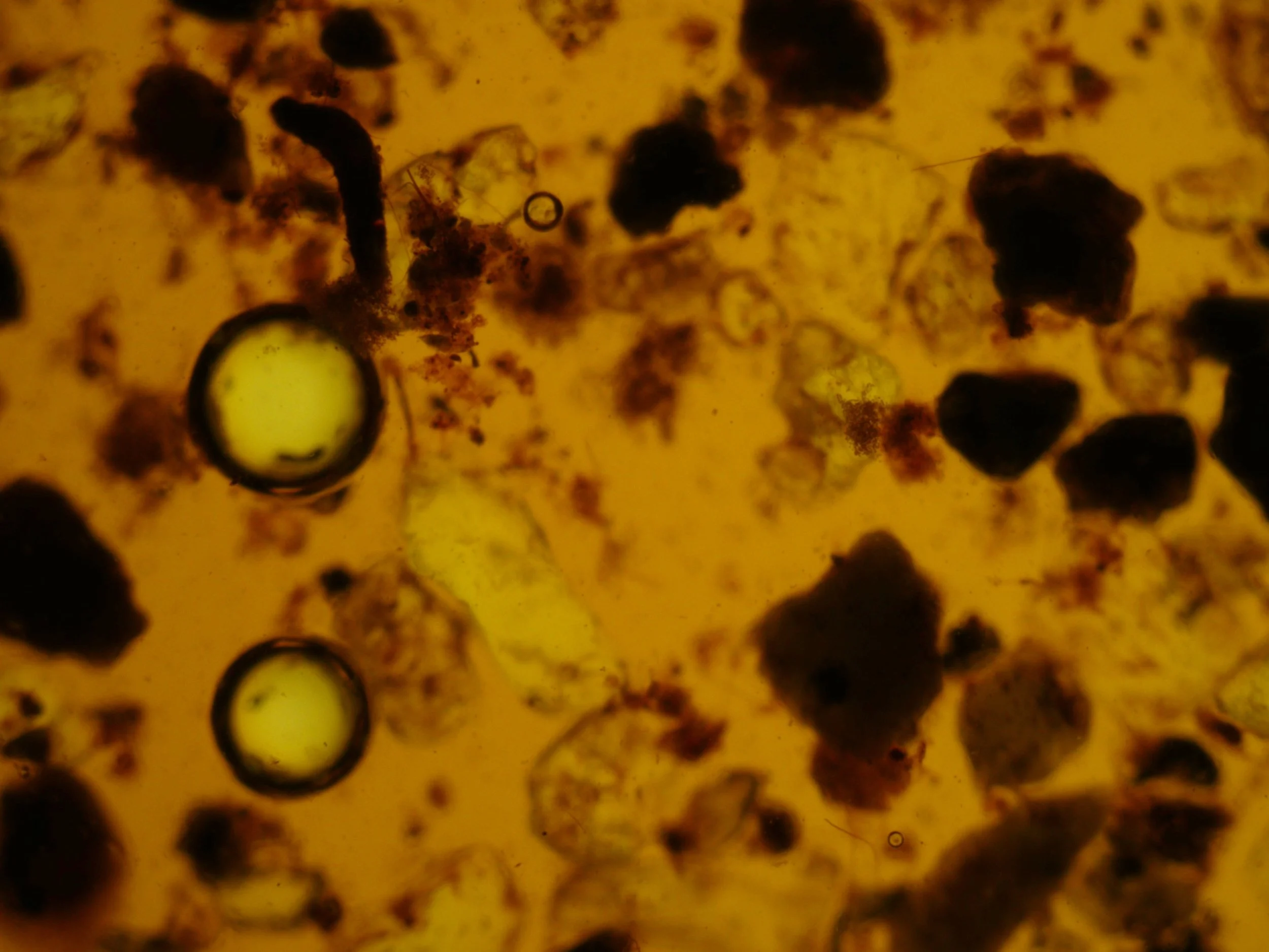
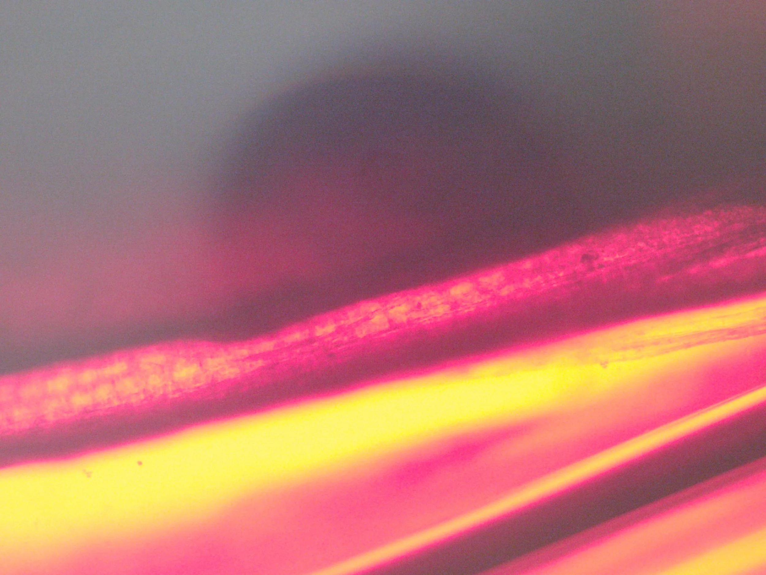
Using Blue Methylene stain…
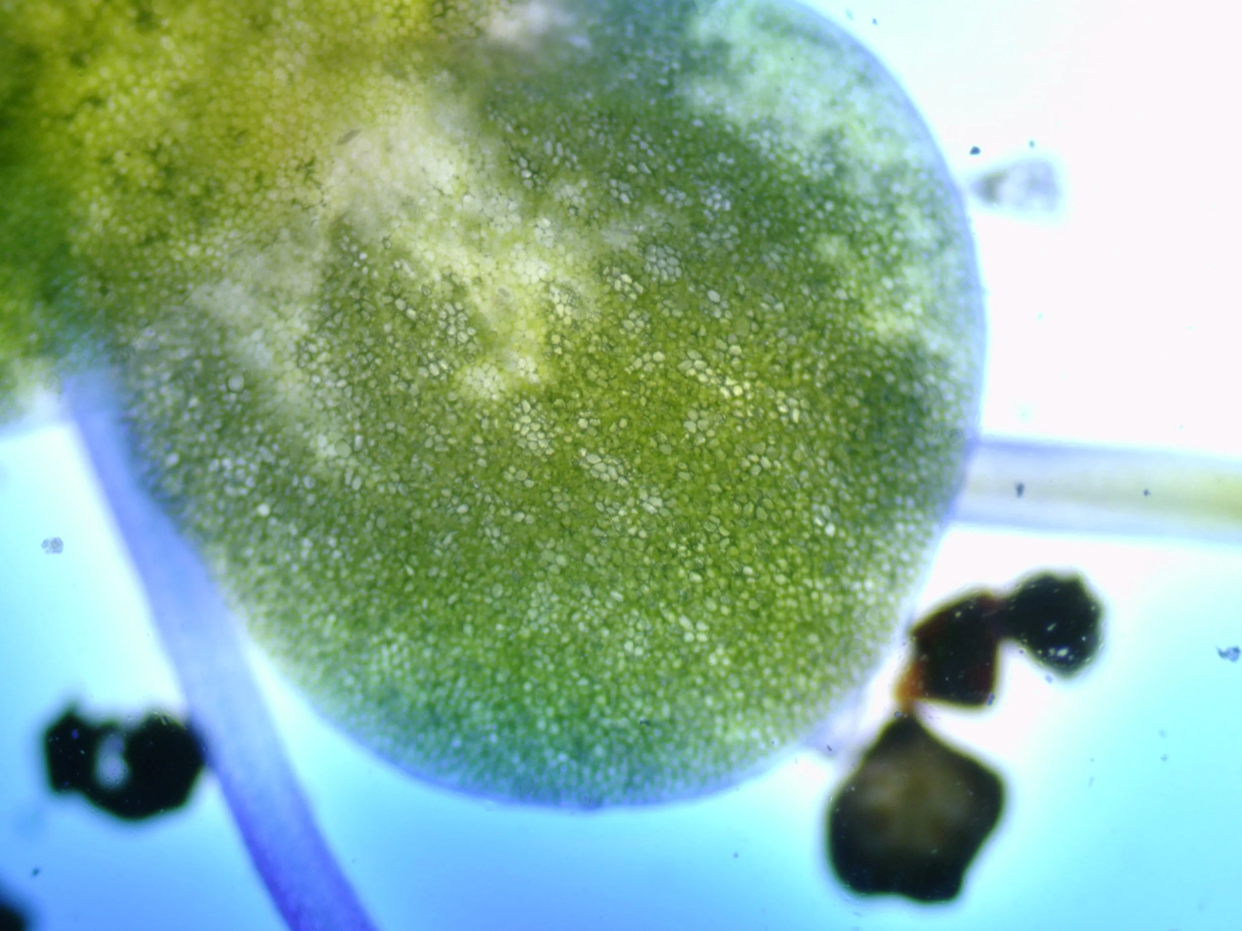
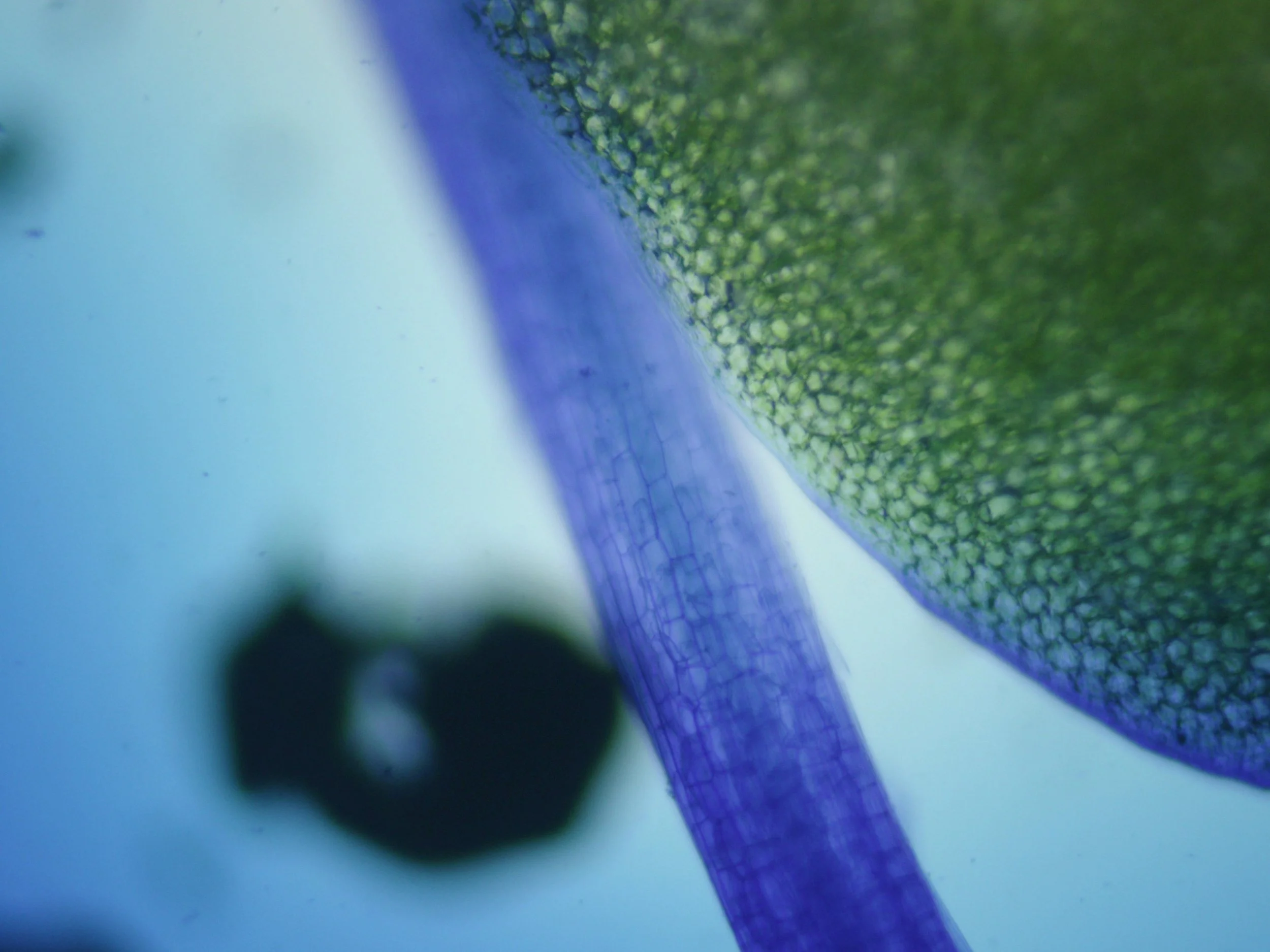
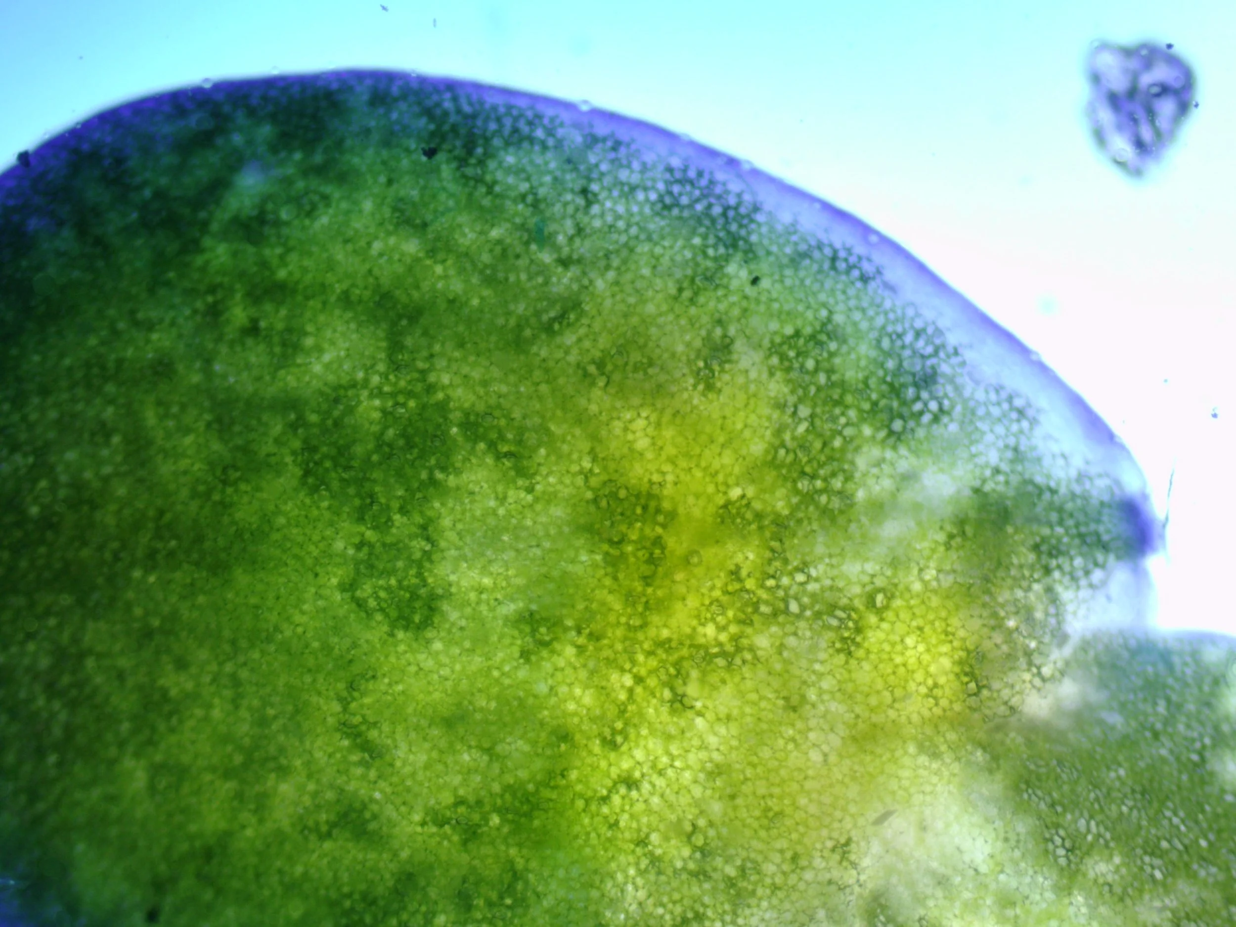
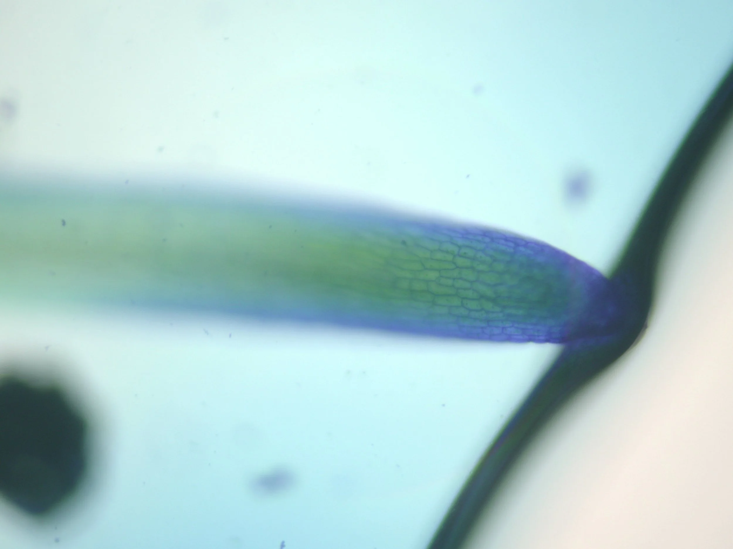
The addition of Methylene Blue produced an unexpected optical transformation. Rather than appearing blue through the camera, areas of the sample shifted into intense pink, magenta and yellow. This is likely to result from the interaction between the stain, the density of the specimen, transmitted microscope light and the camera's colour response, rather than the specimen itself literally becoming pink (The original without the stain is on the far left).