Microscopic Experiments: Staining Rainwater Samples
Currently, I am working in the studio with the microscope and experimenting with microscopy stains, Methylene Blue and Lugol’s Iodine. I decided not to use any of my Lough Neagh samples as I wasn’t one hundred per cent sure what I was doing, to be honest! Instead, I used rainwater that I had collected in a jar over the course of a week.
I wanted to carry out a number of simple experiments: firstly, to capture images of the rainwater sediment under the microscope before staining; secondly, to introduce Methylene Blue and Lugol’s Iodine to the samples; and thirdly, to document how the microscopic images changed over time, photographing them immediately after staining and again after 24 and 36 hours.
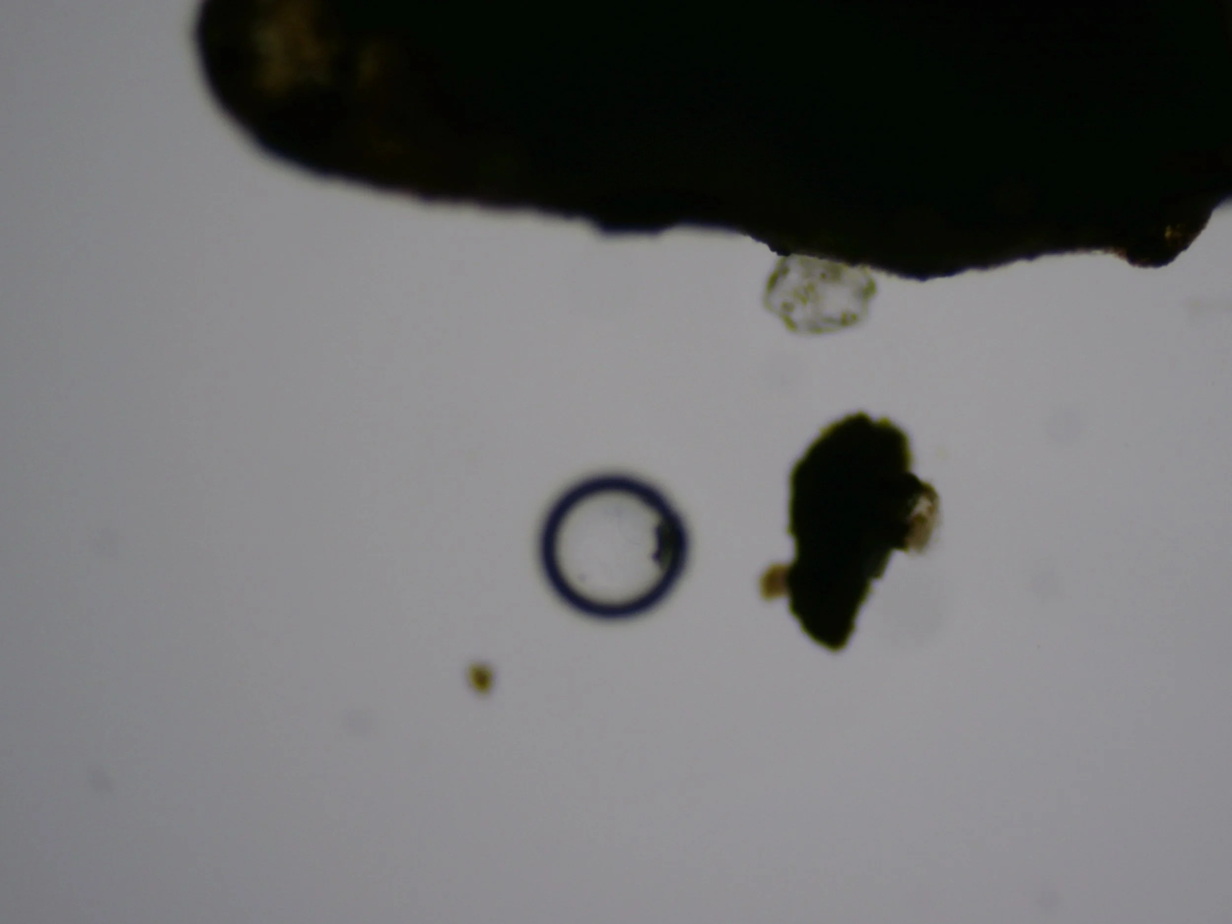
Rainwater sediment under the microscope before staining
The unstained rainwater sample revealed relatively little under the microscope. Although there were small particles and sediment present, visually it was quite sparse. The real change came when I introduced the stains.
Then this happened!!!!!!
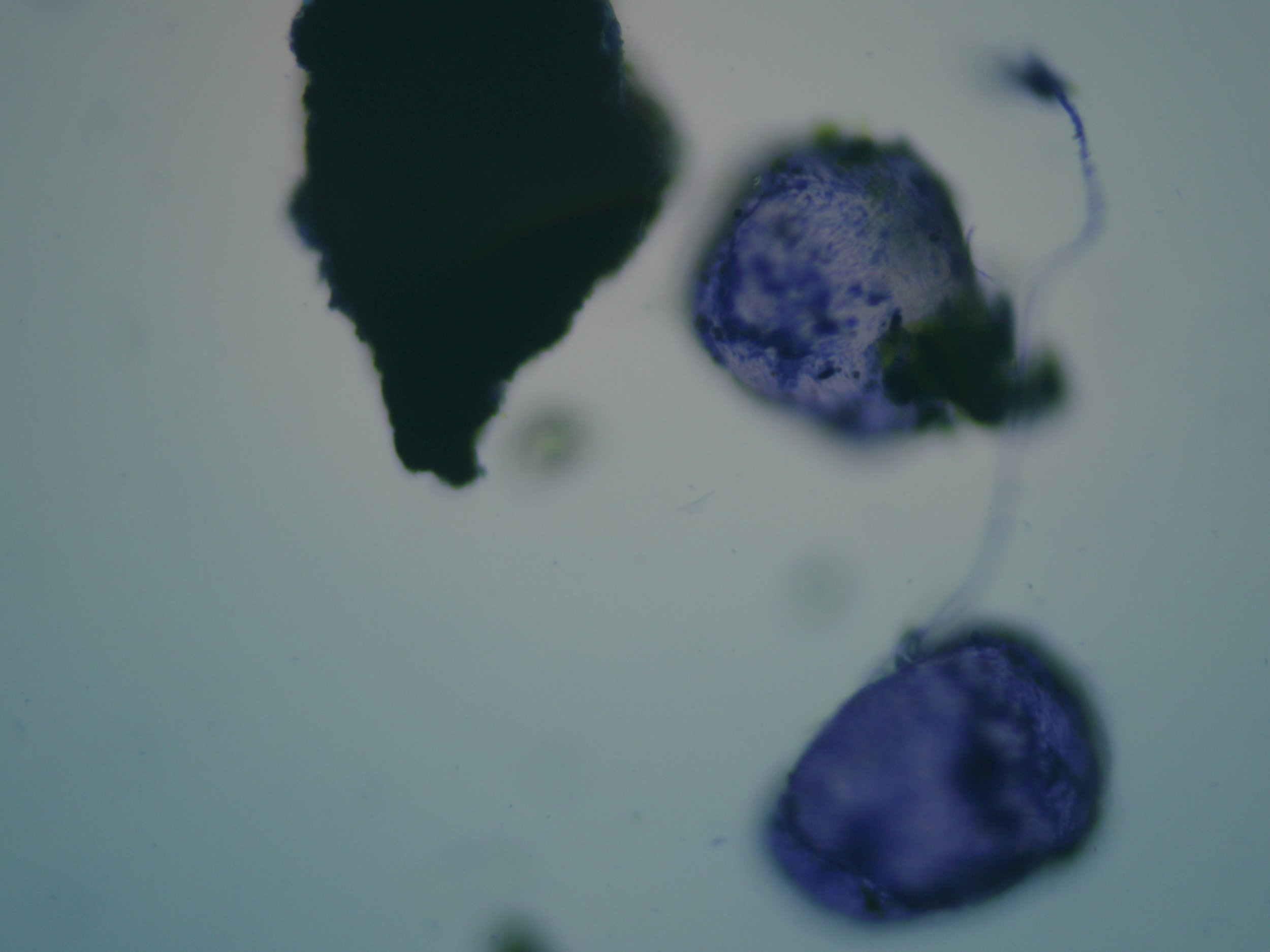
Methylene Blue!!!!! It increases contrast and stains cellular material blue, making otherwise transparent structures easier to see. How fab is this???
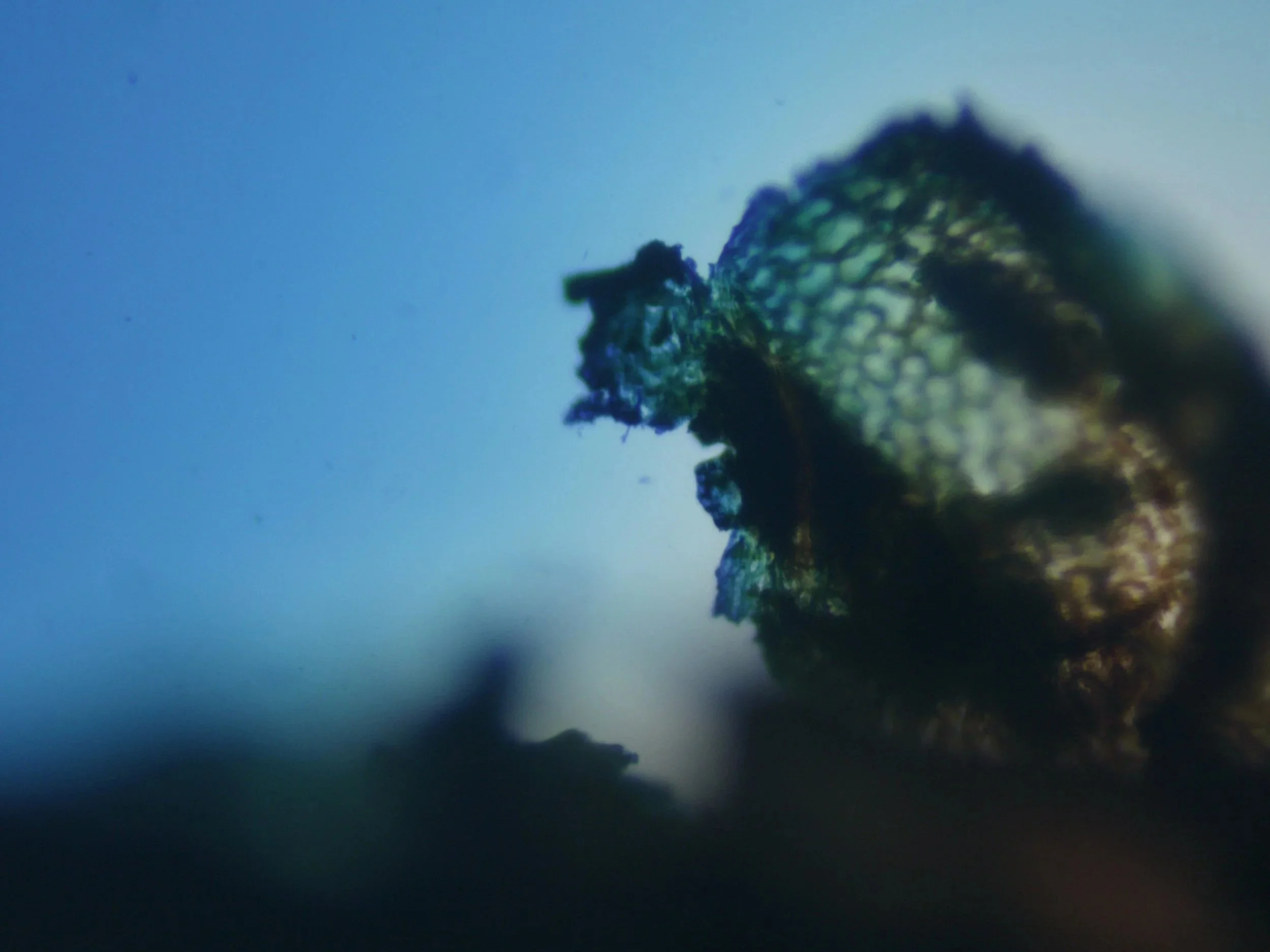
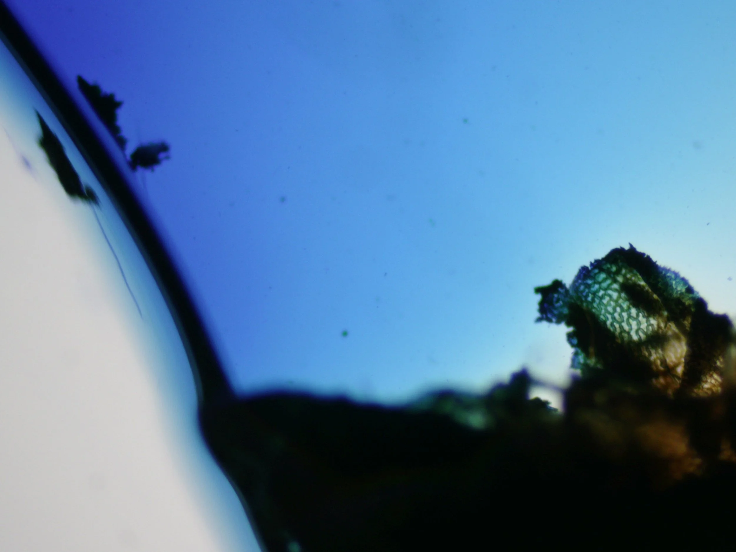
Lugol’s Iodine- equally as fab but a total contrast.
Lugol's stains and preserves algae and other biological material, increasing contrast and producing yellow/brown tones; it also stains starch strongly.
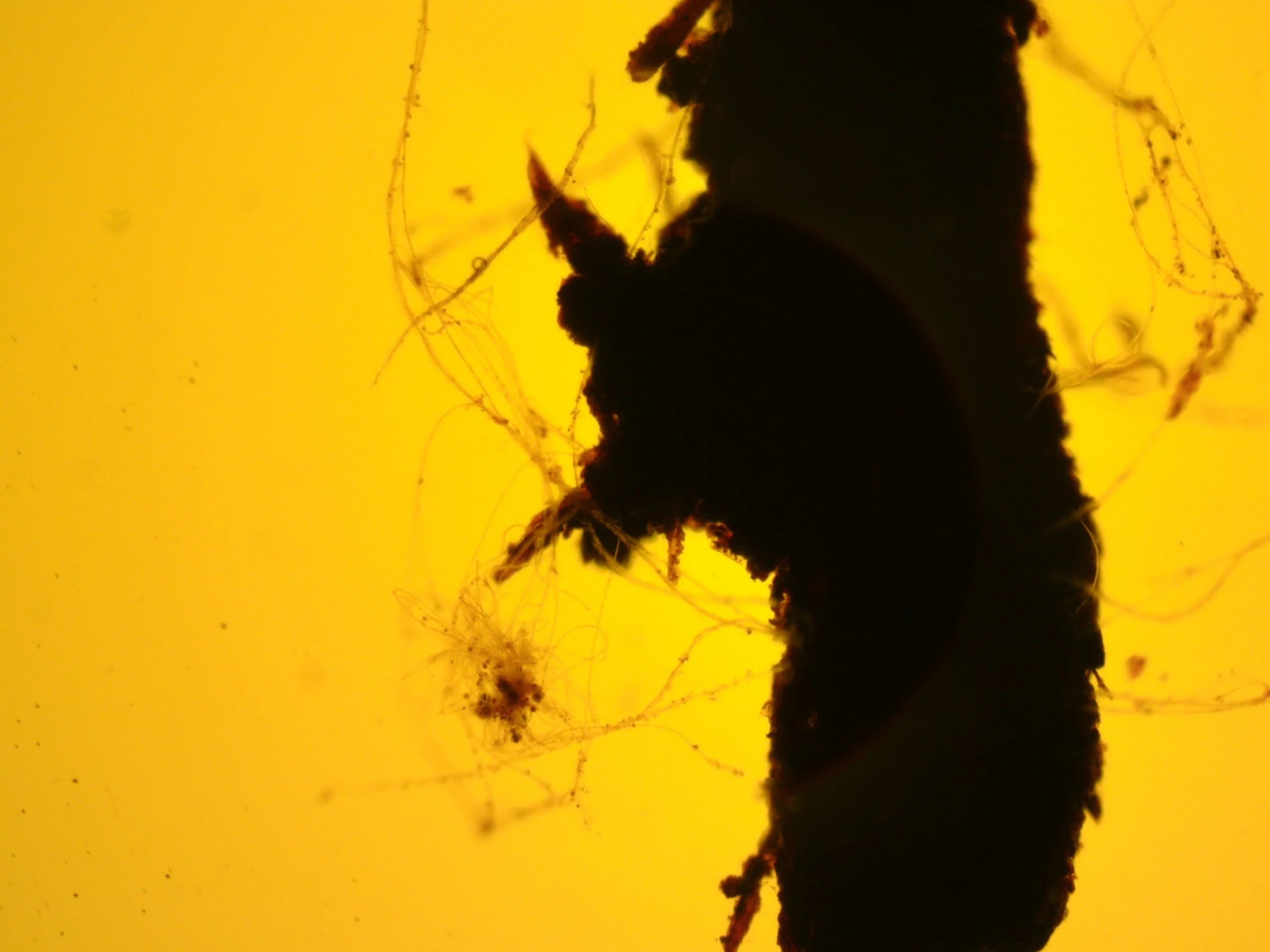
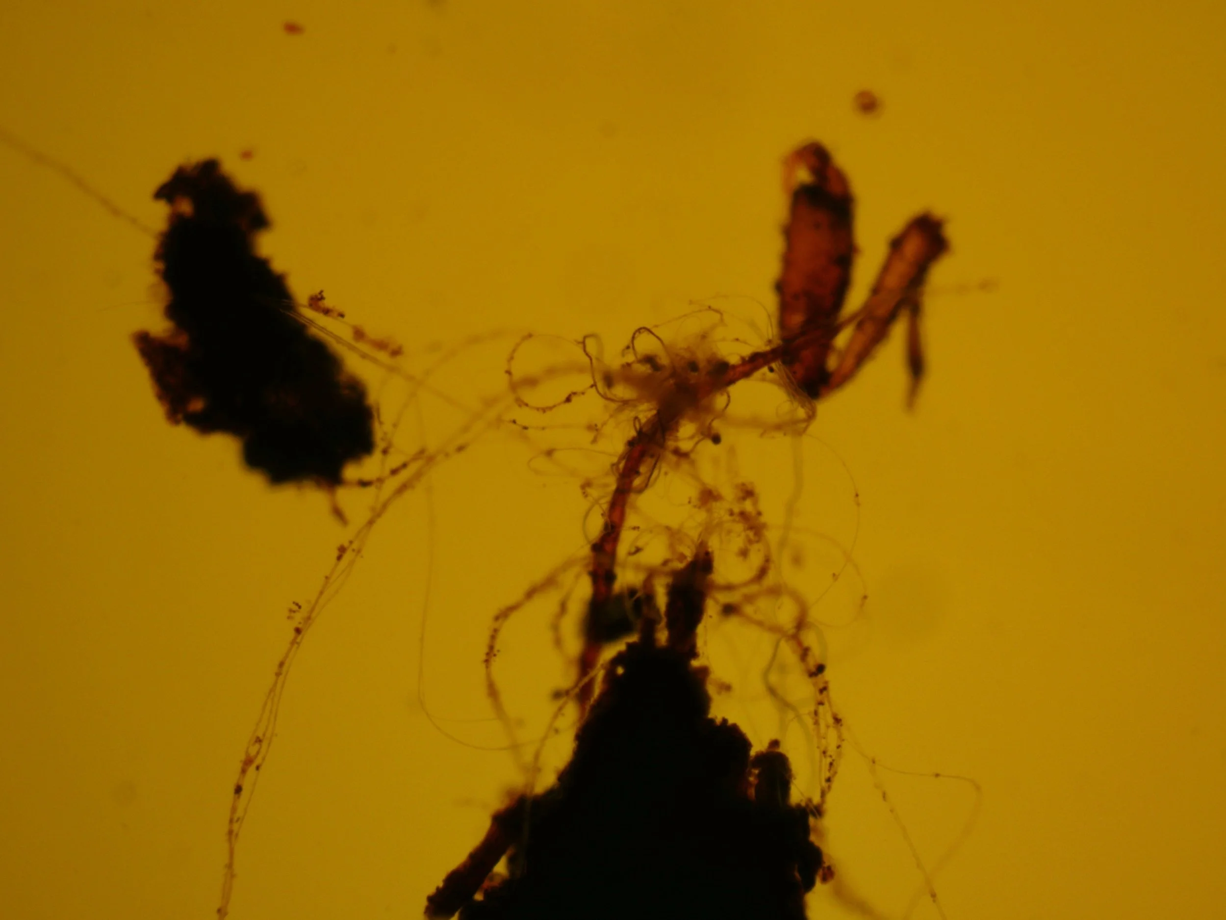
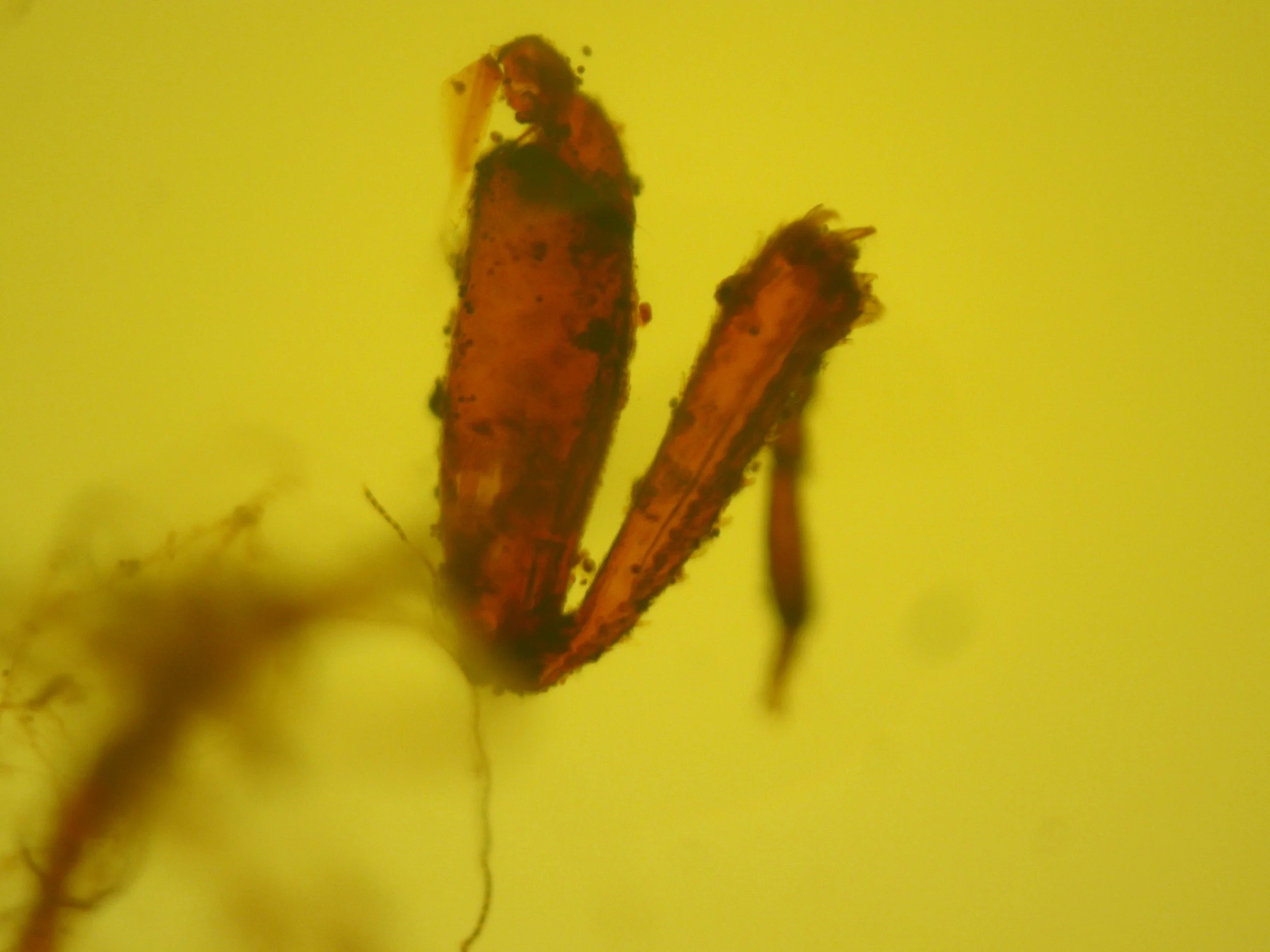
So the stain isn't necessarily revealing something that wasn't physically there before. It's changing the contrast/visibility of material already in the sample AND I JUST LOVE THEM!!! LOL…
I then left the slides overnight and took images of them over 24 and 36 hrs.
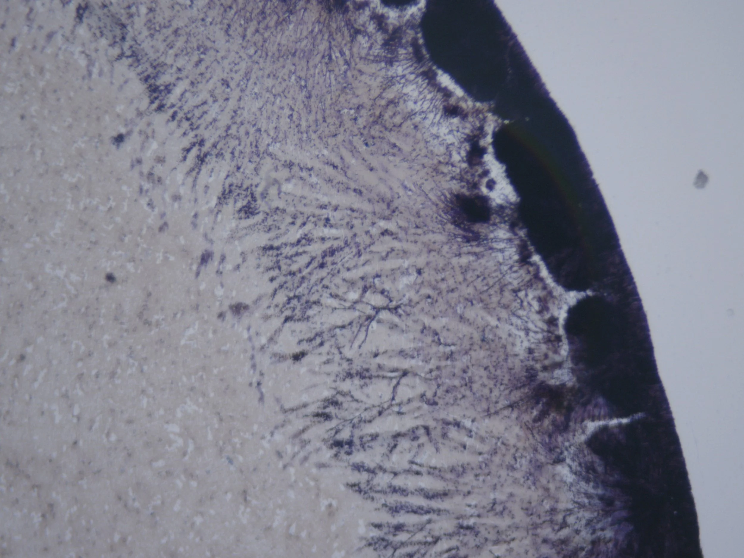
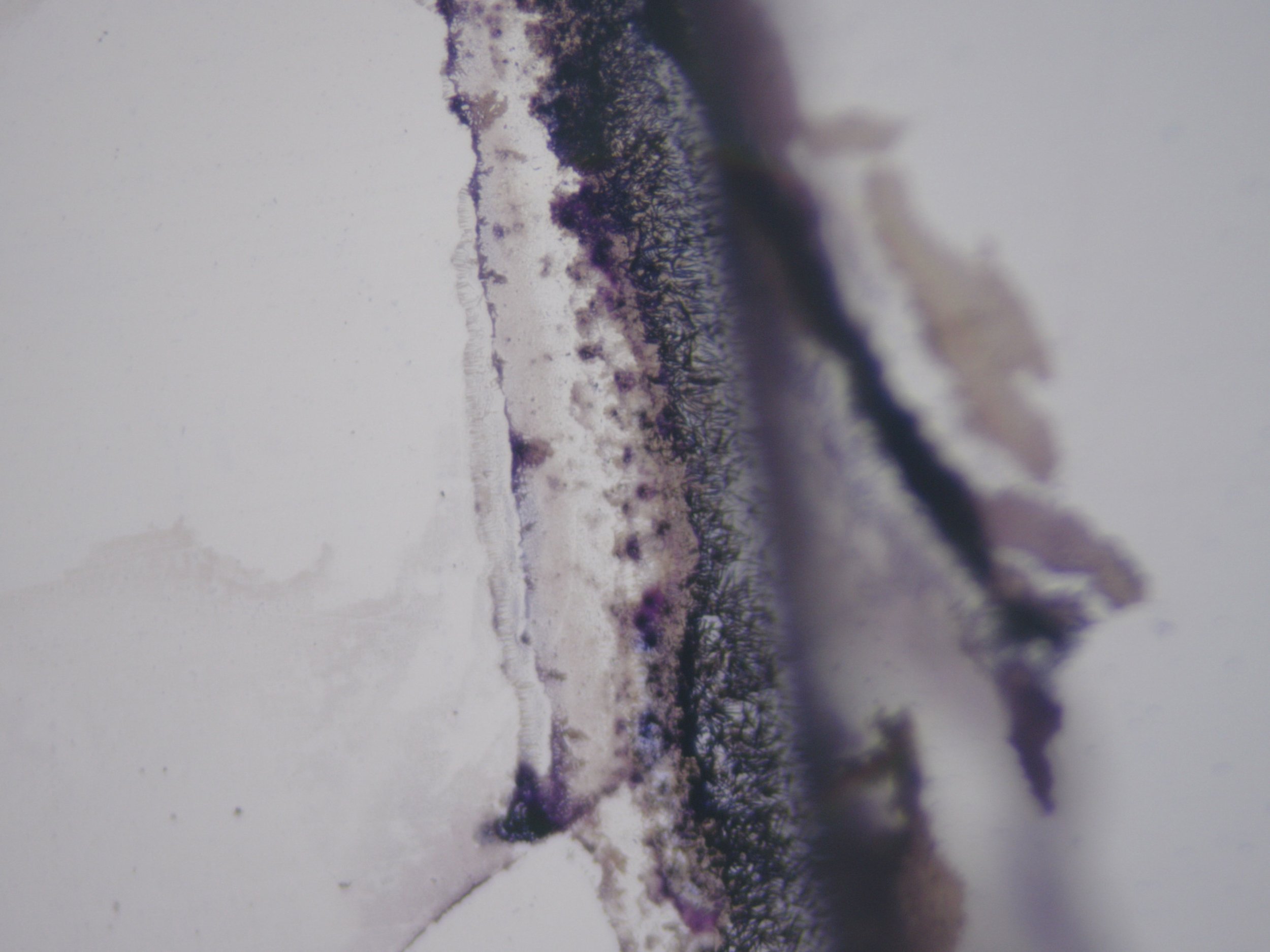
Lugol’s 24 hours after staining: Significant visual change. Colour became less dominant and new translucent, crystalline and branching structures appeared as the sample settled and partially dried beneath the cover slip. Darker concentrations developed around the edges of some areas.
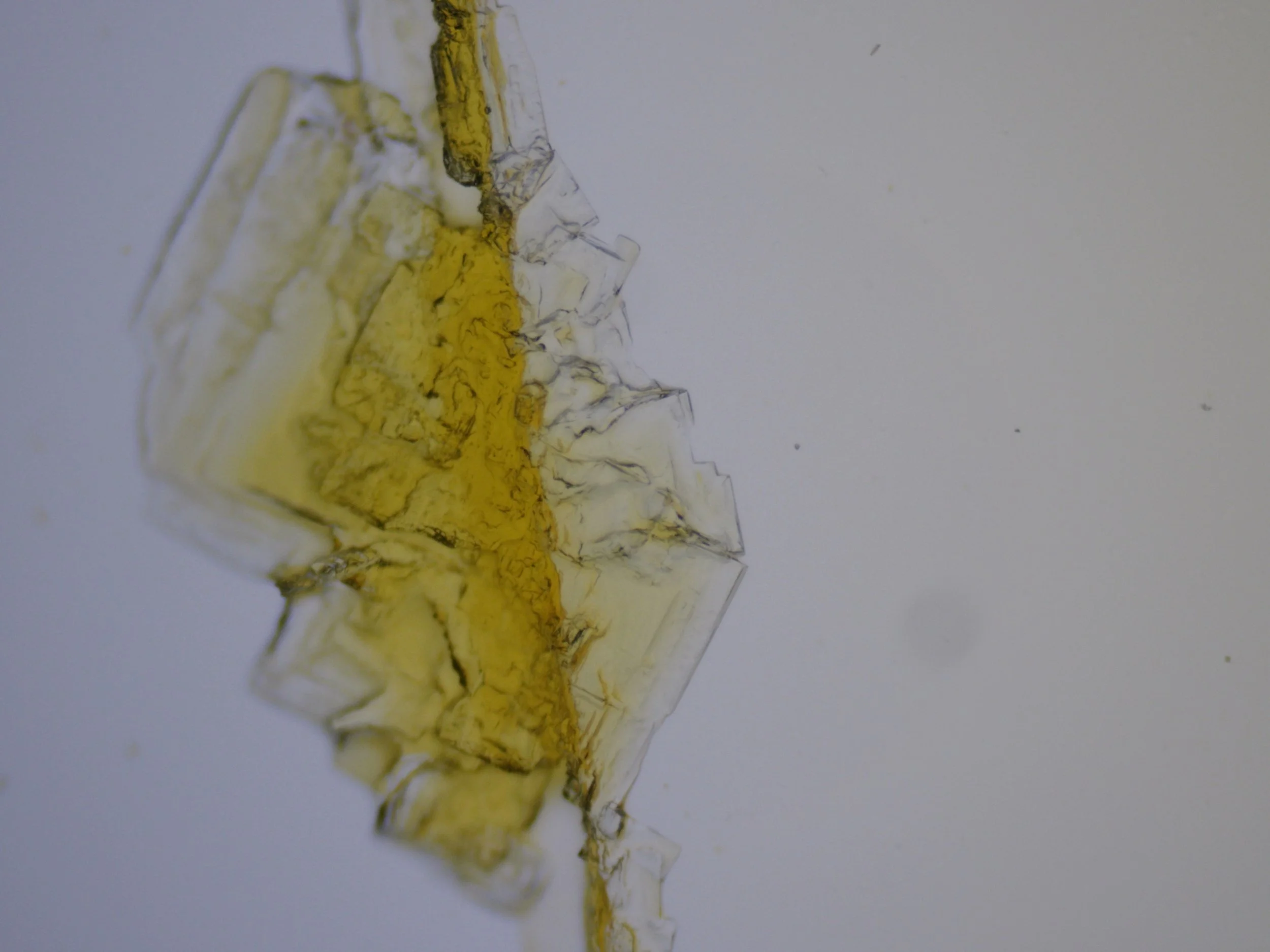
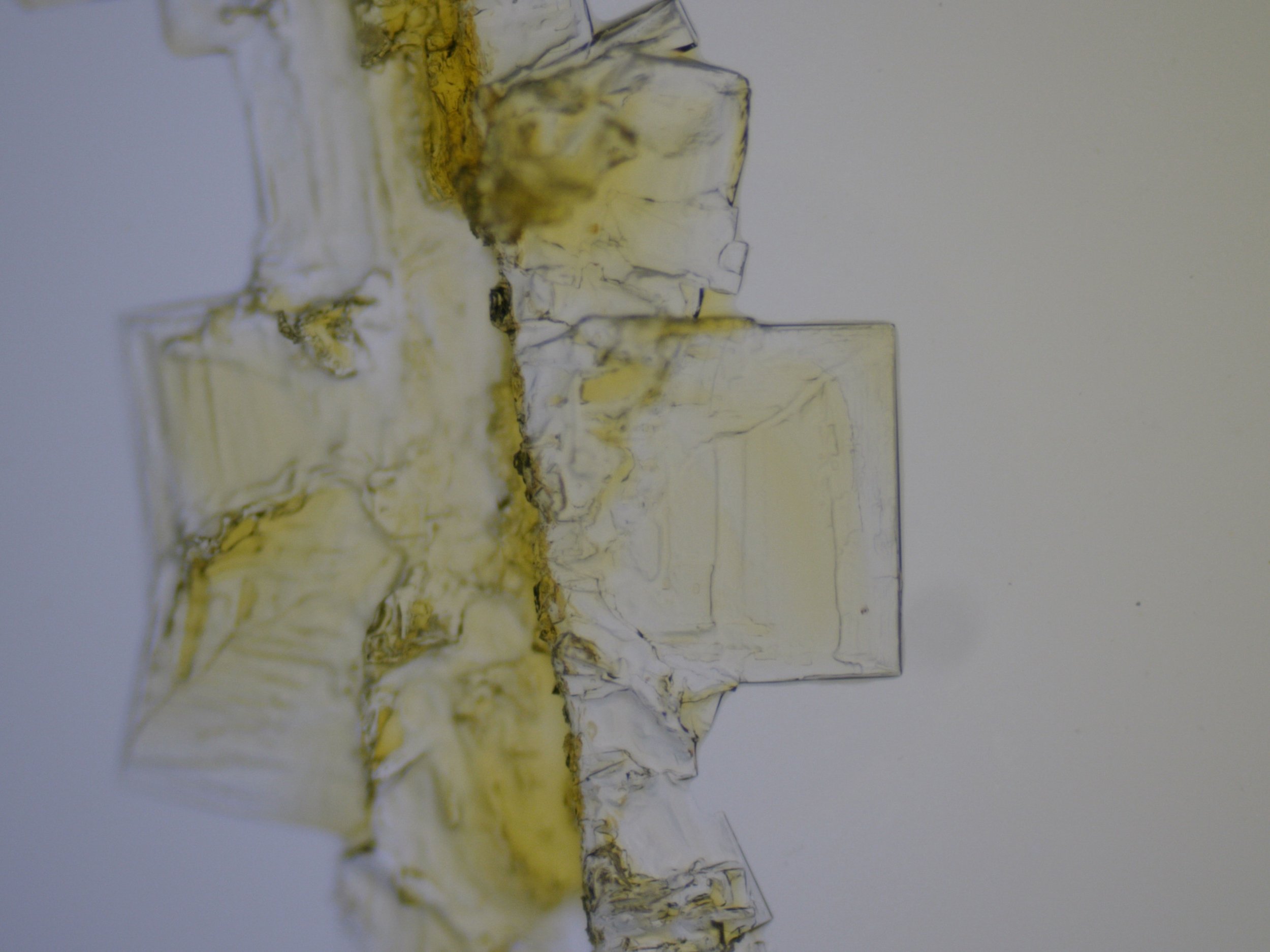
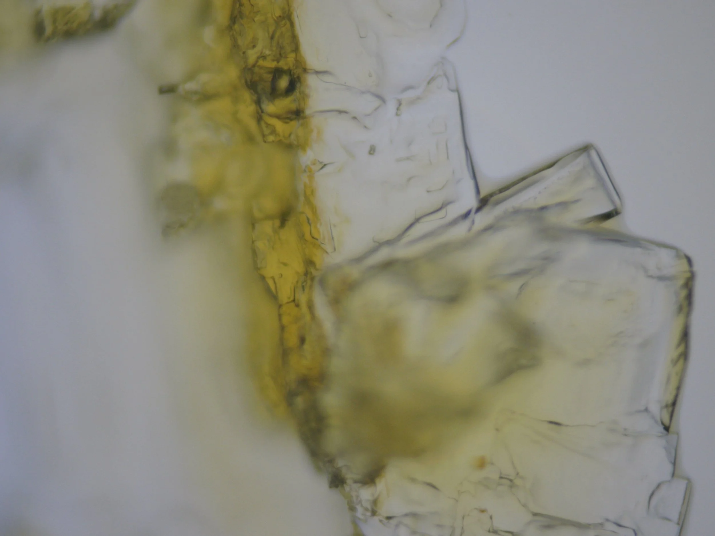
Methylene Blue 24 hrs after Staining:
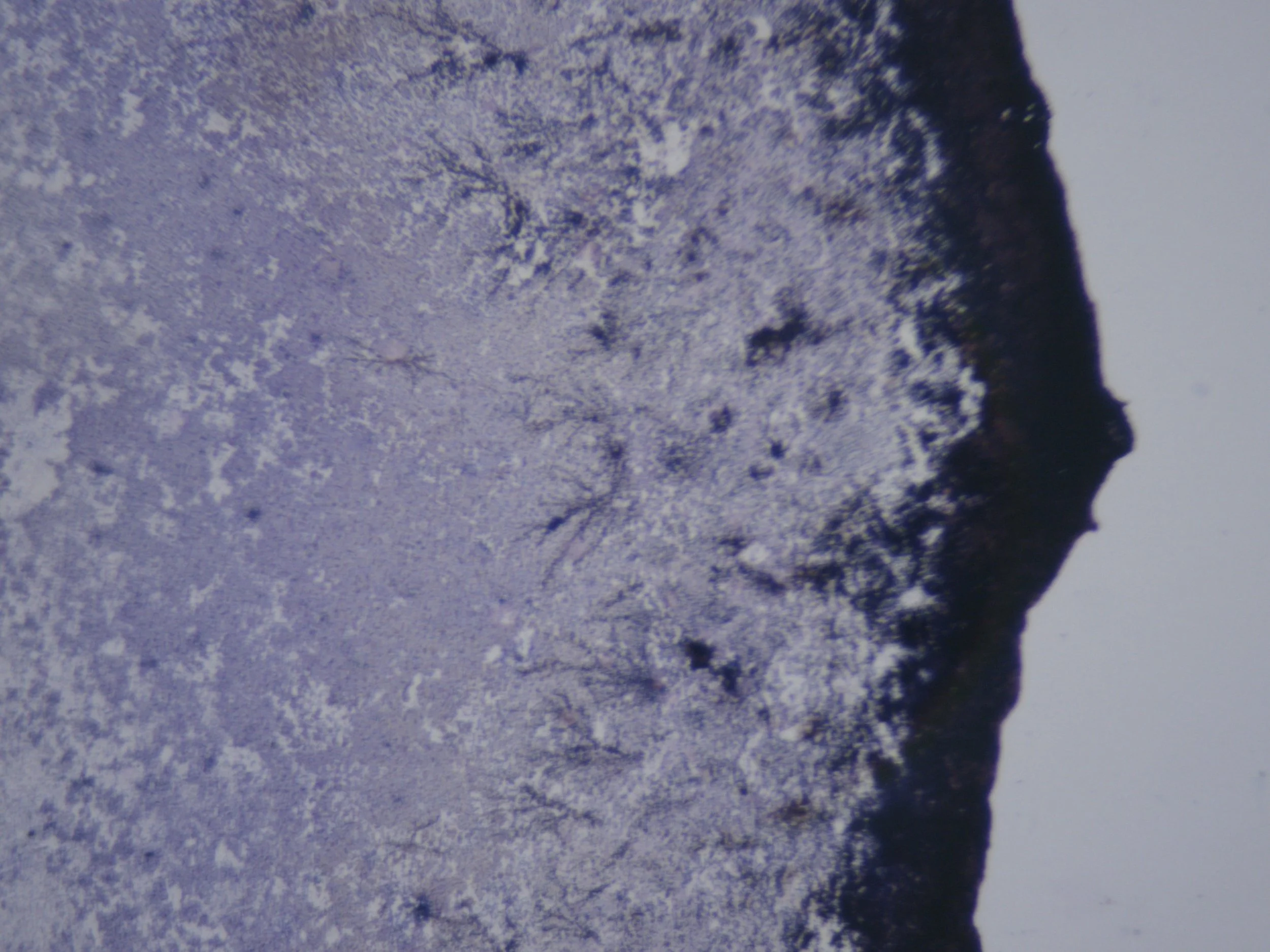
And finally…if you’re still here!!!
36 hrs after staining:
Further changes visible across the slides. The original stain colour has largely dispersed, nearly disappeared except where it settled as spots when I dropped it onto the slide (MBlue).
Translucent angular and branching/crystalline-looking formations have become more pronounced. Dark deposits remain in some areas, with new patterns appearing as the sample continues to settle and dry beneath the cover slip.
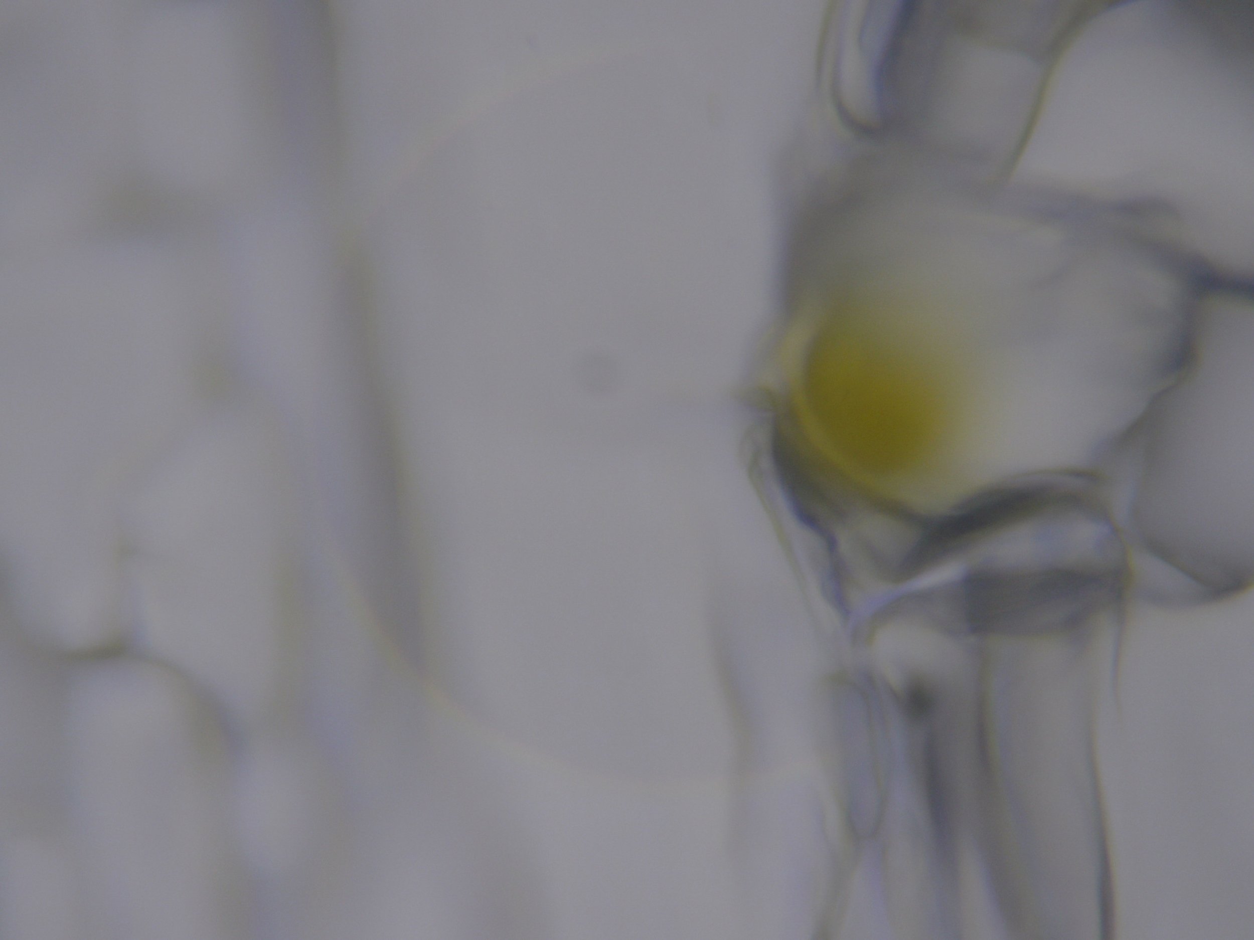
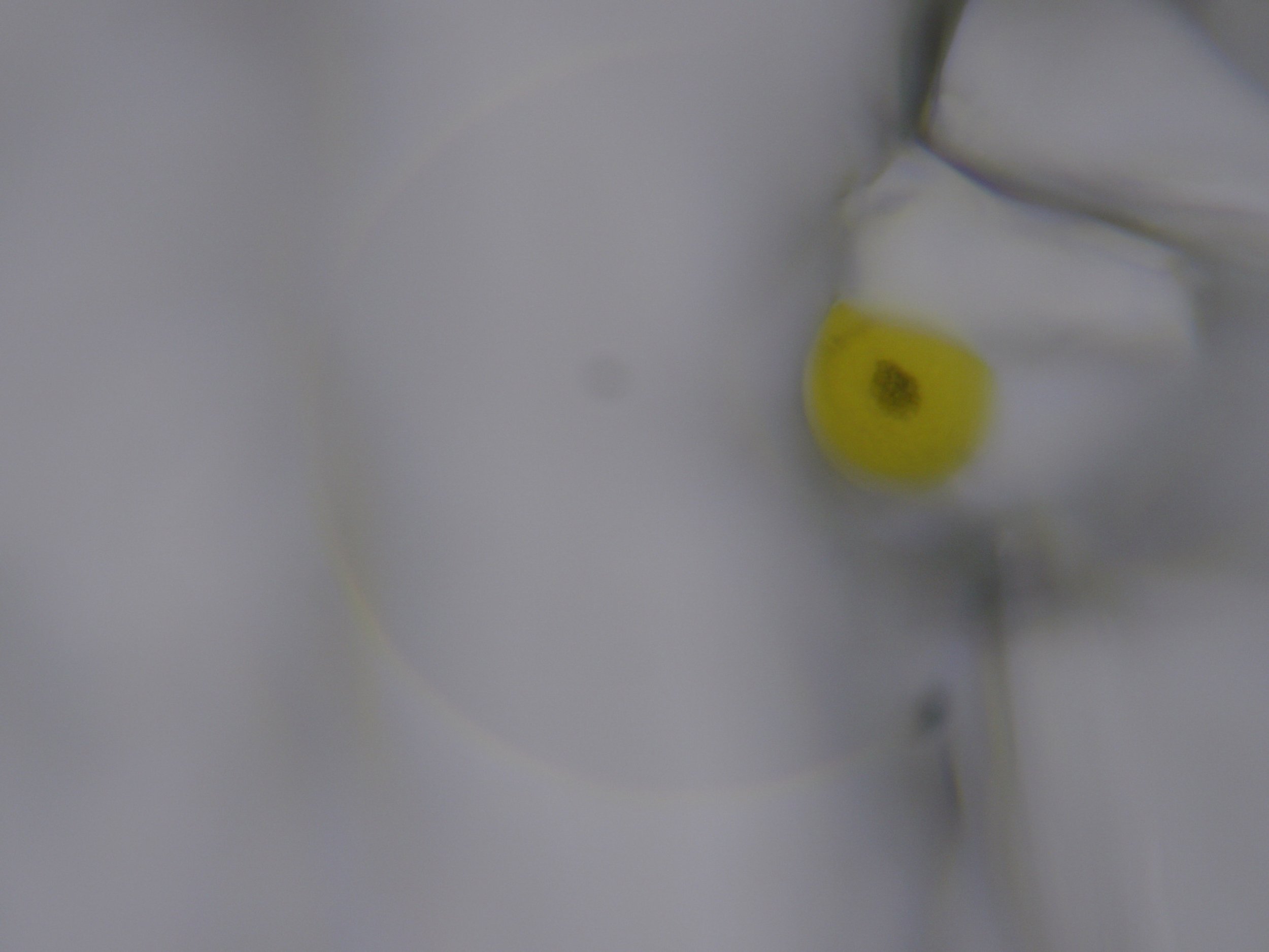
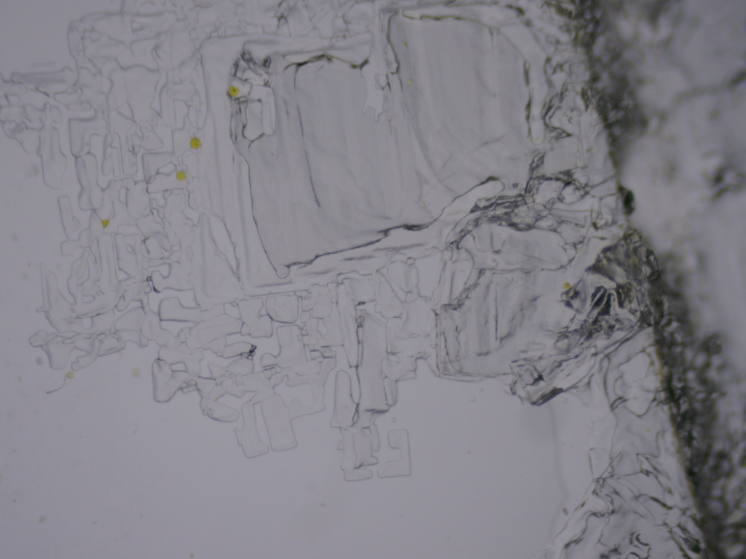
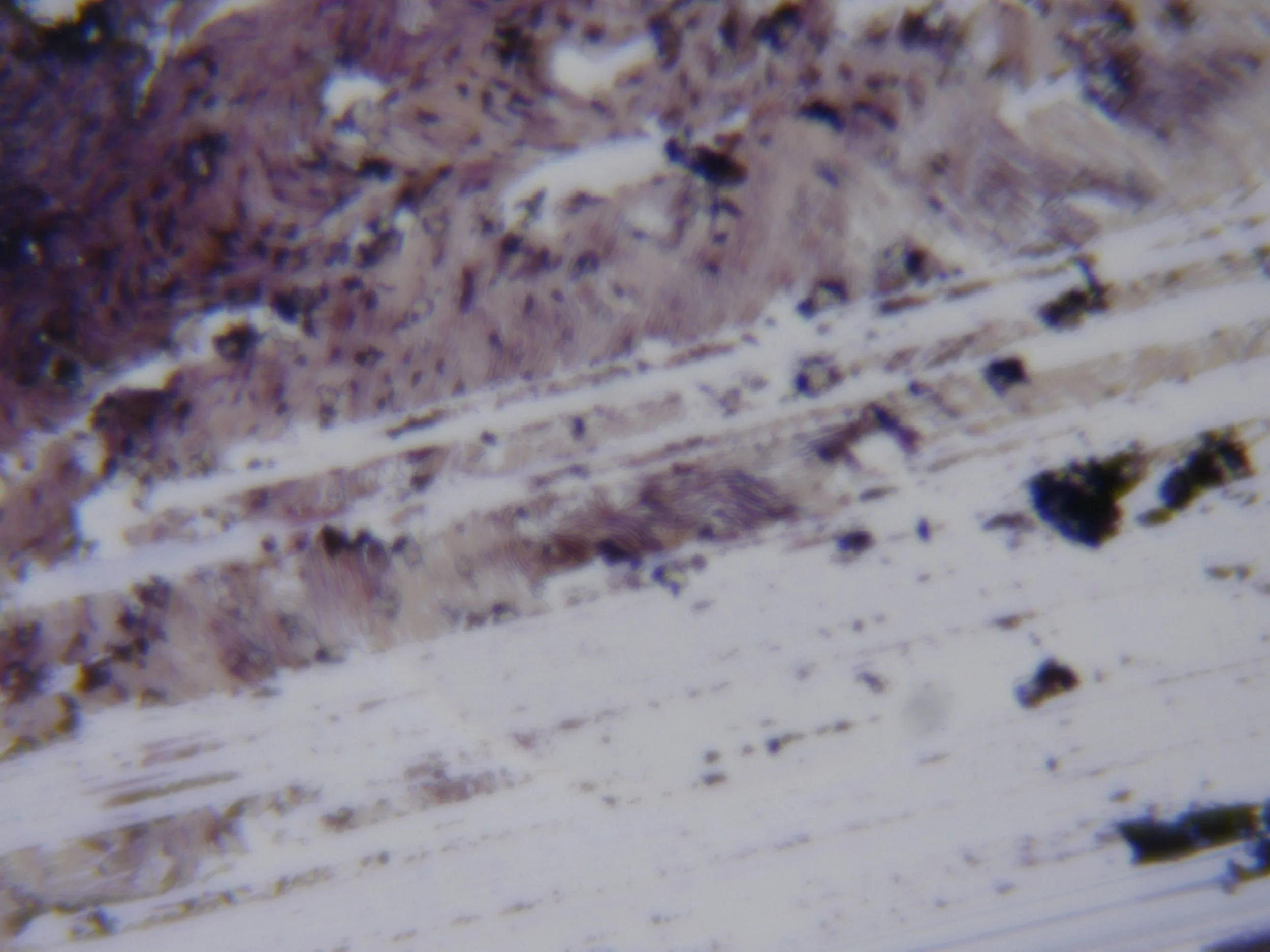
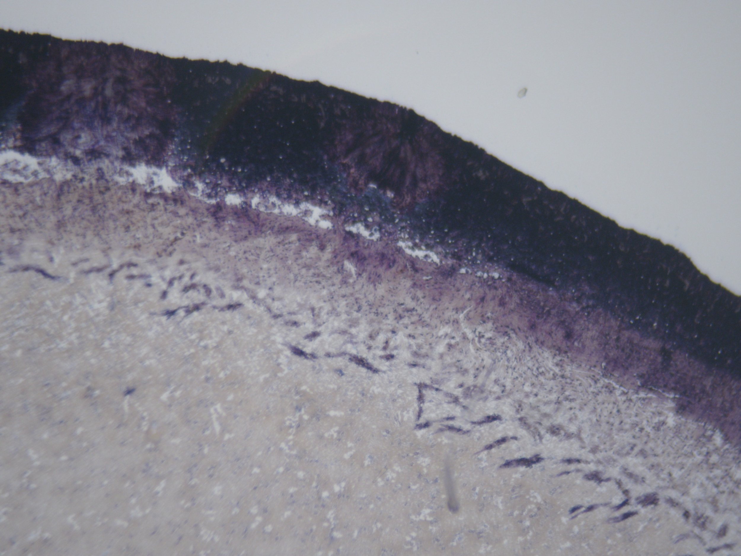
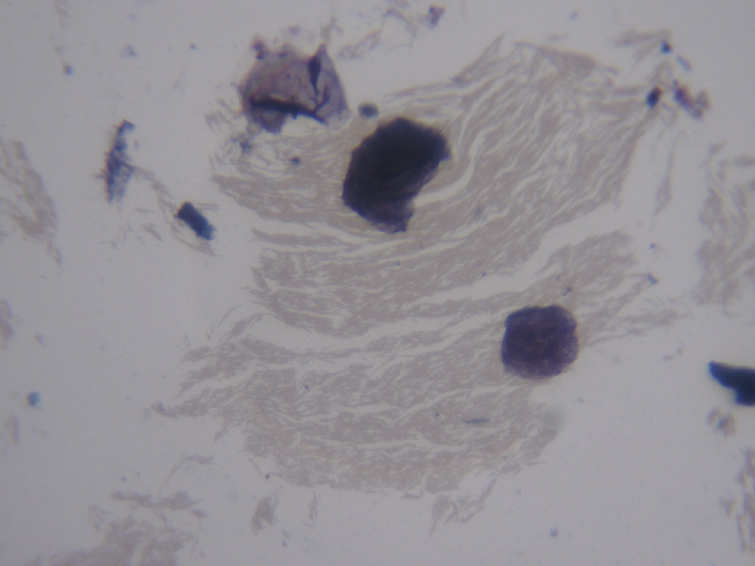
What I was delighted about was that I could actually obtain images from the 40 x lens. 40× images were significantly easier to focus and photograph after the sample had settled/dried beneath the cover slip than when photographing a fresh water droplet.
When I’ve previously been photographing a fresh water droplet, everything is working against you at 40×. The specimen can move in the liquid, the water itself can shift under the cover slip, and you're trying to focus through different depths. At 40×, where your depth of field is already very shallow, even tiny movement can throw the subject out of focus.
With these slides after 36 hours, much of the water has evaporated or redistributed and the material has settled onto the slide/cover slip. Those structures are therefore much more stationary and effectively flatter.
So, every day is a school day peoples…lol…